
Automatic ROI (Regions of Interest) detection method of digital pathologic full slice image

A region of interest and digital pathology technology, applied in character and pattern recognition, instruments, computer parts, etc., can solve problems that cannot be realized, and achieve the effect of easy automatic detection

Inactive Publication Date: 2016-10-12
MOTIC XIAMEN MEDICAL DIAGNOSTICS SYST
View PDF2 Cites 13 Cited by
  • Summary
  • Abstract
  • Description
  • Claims
  • Application Information

AI Technical Summary

Problems solved by technology

For this link, domestic and foreign scholars mainly adopt the method of asking pathologists to mark all the regions of interest in a large number of full slices, using the marked regions as positive samples and the unmarked regions as negative samples for learning. This method has accurate results, but requires It takes a lot of manpower and time for experts, which cannot be realized in large-scale databases

Method used


Image

  • Automatic ROI (Regions of Interest) detection method of digital pathologic full slice image
  • Automatic ROI (Regions of Interest) detection method of digital pathologic full slice image
  • Automatic ROI (Regions of Interest) detection method of digital pathologic full slice image

Examples


Embodiment Construction

[0023] The present invention will be further described below in conjunction with the accompanying drawings and specific embodiments.

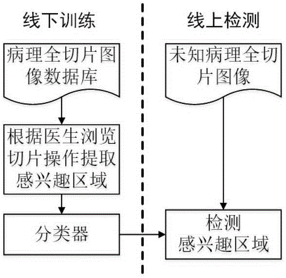
[0024] Such as figure 1 As shown, a method for automatic detection of regions of interest in digital pathological full-slice images is applied to the digital pathological full-slice image database, including the offline training stage and the online detection stage.

[0025] (1) Operation steps in the offline training phase

[0026] (1) The viewport of interest in each full slice in the database is extracted through the standard operation set by the doctor to observe the full slice

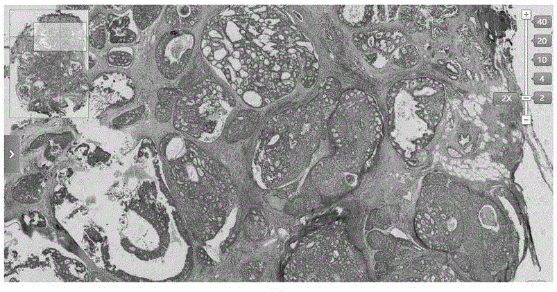
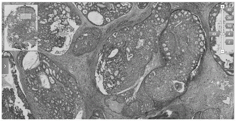
[0027] The present invention obtains the region of interest through the doctor's viewport record, and the viewport refers to the visible part of the doctor's full slice on the screen when observing, such as figure 2 , image 3 , Figure 4 As shown in , the upper left corner is the thumbnail of the full slice and the position of the viewport in the full slice, ...


PUM


Abstract

The invention discloses an automatic ROI detection method of a digital pathologic full slice image. The method is applied to a digital pathologic full slice image database, and comprises an offline training stage and an online detection stage. A doctor observes full slices to set an ROI viewport in each full slice in a standard extraction database, the ROI viewpoints are spliced to obtain a complete ROI, an overlapped slide window method is used to extract a rectangular area from the ROI, the rectangular area serves as a positive sample, a rectangular area collected from a non ROI serves as a negative sample, and the samples are used to train a classifier; and the classifier is used to determine a rectangular area extracted from unknown full slices and return a result. According to the invention, rectangular area samples can be obtained by only recording a slice observing operation of the doctor, the sample obtaining difficulty can be greatly reduced on the premise that the correctness is ensured, automatic detection for full slice ROI is easy to realize, time and labor are saved, and detection is rapid and accurate.

Description

technical field [0001] The invention relates to the fields of digital image processing and machine learning, in particular to an automatic detection method for a region of interest in a digital pathological full slice image. Background technique [0002] Digital pathological full slide image (hereinafter referred to as full slide) is a large-size, high-resolution digital image obtained by scanning and collecting traditional glass pathological slides through a fully automatic microscope or an optical magnification system. It is an important basis for pathologists in diagnosis. In recent years, with the rapid development of pathology and computer technology, the number of whole slides has increased rapidly. Automatic detection of regions of interest in unknown full slices through machine learning through a large number of known full slices is of great reference value for rapid diagnosis by pathologists and automatic computer diagnosis. [0003] In this process, the acquisitio...

Claims


Application Information

Patent Timeline
IPC IPC(8): G06K9/32G06K9/62
CPCG06V10/25G06F18/2411
Inventor 姜志国麻义兵
Owner MOTIC XIAMEN MEDICAL DIAGNOSTICS SYST
Learn More