Method for segmenting aorta in CT image combining edge and area characteristics

A CT image and regional feature technology, applied in the field of medical image processing, can solve problems such as unsatisfactory effects

Active Publication Date: 2016-10-26
福建省公田软件股份有限公司
View PDF4 Cites 13 Cited by
  • Summary
  • Abstract
  • Description
  • Claims
  • Application Information

AI Technical Summary

Problems solved by technology

This method can accurately segment the complete aorta with high definition and less noise, but once the image is noisy or aortic dissection occurs, the effect is not ideal
The Fourth People's Hospital of Shenyang disclosed a method for segmenting blood vessels in fundus images based on an extensible active contour model (patent application number CN201510274619, publication number CN104867151A), which was applied for by the Fourth People's Hospital of Shenyang. The method of constructing the direction field by feature and using the ductile active contour segmentation model to segment the fundus blood vessels, this method takes advantage of the characteristics of the blood vessels, but is not suitable for the segmentation of the aorta

Method used


Image

  • Method for segmenting aorta in CT image combining edge and area characteristics
  • Method for segmenting aorta in CT image combining edge and area characteristics
  • Method for segmenting aorta in CT image combining edge and area characteristics

Examples


Embodiment Construction

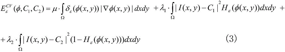
[0053] The present invention is a segmentation method of aorta in a CT image that combines edge and region features. The edge features of the segmented image are integrated into the traditional CV level set model, and a circle is added as a priori energy item to construct a new energy function E. Its specific implementation steps are as follows:

[0054] Step 1. Construct the energy function E of the traditional CV model:

[0055] The CV model is a typical region-based level set model. The segmented image I(x,y) whose domain of definition is Ω is divided into two homogeneous regions by a closed contour line C, that is, the inside and outside of the closed contour line C , the average gray value of each region is C 1 and C 2 , the energy functional E(C) can be expressed as follows:

[0056]

[0057] In the above formula, x and y respectively represent the horizontal and vertical coordinates of the pixel in the image;

[0058] If the closed contour line C is located at th...


PUM


Abstract

The invention discloses a method for segmenting aorta in a CT image combining edge and area characteristics. The method combines edge and area characteristics, by means of level set theory, can obtain clear and accurate object contour, implements segmentation of the aorta of the CT image, has very rapid operation and provides accurate segmentation result, better addresses the extraction of the aorta in the CT image, and provides a reference to medical diagnosis.

Description

technical field [0001] The invention belongs to the technical field of medical image processing, and in particular relates to a method for segmenting aorta in a CT image based on a level set method and fusing image edge and region features. Background technique [0002] Image segmentation is a key technology in image processing. CT (Computed Tomography) can provide high-resolution and high-quality CT images. CT images accurately describe the anatomical structure and function of medical tissues and organs. Extracting the doctor's interested area in the CT image can facilitate the doctor's clinical diagnosis and surgical treatment. Different from general network images, CT images have complex imaging mechanisms and special data. Some traditional segmentation methods can no longer meet the needs of image segmentation. [0003] Although there are many studies on medical image segmentation, a universal segmentation method has not been found. For the segmentation of aorta in C...

Claims


Application Information

Patent Timeline
Patent Type & Authority Applications(China)
IPC IPC(8): G06T7/00
CPCG06T7/0012G06T2207/10081G06T2207/30101
Inventor 骆炎民杨珺柳培忠
Owner 福建省公田软件股份有限公司
Why Eureka
Try Eureka