Liver and kidney medical image data co-processing system

A data processing and medical image technology, applied in the field of medical image processing systems, can solve the problems of unable to process three-dimensional CT images of liver and kidney at the same time, unable to interactively display three-dimensional CT images of liver, etc.

Active Publication Date: 2016-11-09
THE AFFILIATED HOSPITAL OF QINGDAO UNIV
View PDF11 Cites 5 Cited by
  • Summary
  • Abstract
  • Description
  • Claims
  • Application Information

AI Technical Summary

Problems solved by technology

However, the 3D reconstruction method of liver CT images cannot interactively display 3D CT stereo images of the liver, and cannot simultaneously process liver and kidney data to generate 3D CT stereo images

Method used


Image

  • Liver and kidney medical image data co-processing system

Examples


Embodiment Construction

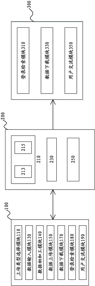
[0057] Please refer to figure 1 According to Embodiment 1 of the present invention, a liver and kidney medical image data collaborative processing system is provided, including: several uploading user terminals 100 , a data processing center 200 and several downloading user terminals 300 . Several uploading user terminals 100 are connected to the data processing center 200 through the Internet to upload initial data to the data processing center 200 . The data processing center 200 is used to generate a three-dimensional image of the liver and a three-dimensional image of the kidney. Several downloading user terminals 300 are connected to the data processing center 200 through the Internet to download the three-dimensional images of the liver or the three-dimensional images of the kidneys of the data processing center 200 .

[0058] Each upload user terminal 100 includes an upload type selection module 110 , a data input module 130 , a data preliminary processing module 140 a...


PUM


Abstract

The invention discloses a liver and kidney medical image data co-processing system which comprises a data processing center, a plurality of uploading user terminals and a plurality of downloading user terminals, wherein the plurality of uploading user terminals are connected with the data processing center through the Internet to upload initial data to the data processing center, and the plurality of downloading user terminals are connected with the data processing center through the Internet to download a liver or kidney three-dimensional image of the data processing center. The data processing center at least comprises an information database and a three-dimensional image processing module used for generating a specific liver three-dimensional image based on a liver CT image and generating a specific kidney three-dimensional image based on a kidney CT image, wherein the information database is used for classifying and storing the specific liver three-dimensional image from the three-dimensional image processing module according to specific liver source information and classifying and storing the specific kidney three-dimensional image from the three-dimensional image processing module according to specific kidney source information.

Description

technical field [0001] The invention relates to a medical image processing system, in particular to a medical image three-dimensional visualization system. Background technique [0002] The 3D reconstruction technology of medical images can make full use of medical image volume data such as CT and MRI, and use surface rendering or volume rendering imaging algorithms to obtain 3D projection images from any perspective perspective as needed, which is convenient for doctors to observe the internal structure of the human body. Using 3D reconstruction technology to process medical images, construct 3D models, and project and display the 3D models from different directions to extract information about relevant organs, so that doctors can obtain quantitative descriptions of the size, shape and spatial position of organs of interest. 3D reconstruction technology enables doctors to observe the 3D structure of organs intuitively and quantitatively, and enhance various original details...

Claims


Application Information

Patent Timeline
Patent Type & Authority Applications(China)
IPC IPC(8): G06F19/00G06F17/30H04L29/08G06T7/00G06T15/00G06T5/00
CPCH04L67/06G06F16/50G06T5/002G06T5/003G06T15/005G06T2207/20032G06T2207/30056G06T2207/10081
Inventor 魏宾董蒨朱呈瞻董冰子周显军
Owner THE AFFILIATED HOSPITAL OF QINGDAO UNIV
Why Eureka
Try Eureka