Gastrointestinal tumor microscopic hyper-spectral image processing method based on convolutional neural network

A convolutional neural network and hyperspectral image technology, applied in the field of gastrointestinal tumor microscopic hyperspectral image processing based on convolutional neural network, can solve the problem of heavy workload for doctors, difficult quantitative analysis, strong subjectivity of visual observation, etc. question

Inactive Publication Date: 2016-11-09
SHANDONG UNIV
View PDF0 Cites 43 Cited by
  • Summary
  • Abstract
  • Description
  • Claims
  • Application Information

AI Technical Summary

Problems solved by technology

A large number of medical images require doctors to make judgments through visual observation. On the one hand, it causes a he...

Method used


Image

  • Gastrointestinal tumor microscopic hyper-spectral image processing method based on convolutional neural network
  • Gastrointestinal tumor microscopic hyper-spectral image processing method based on convolutional neural network
  • Gastrointestinal tumor microscopic hyper-spectral image processing method based on convolutional neural network

Examples


Embodiment Construction

[0053]The present invention is described in detail below in conjunction with accompanying drawing:

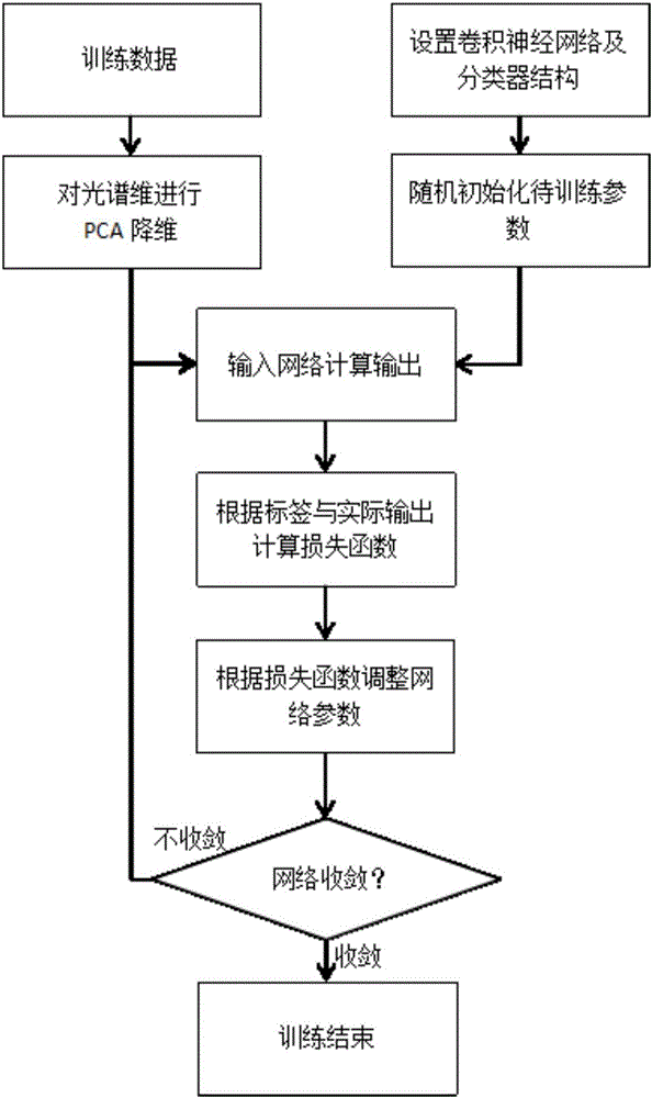
[0054] The network training method in the microscopic hyperspectral image detection of gastrointestinal tumors based on convolutional neural network includes the following steps:

[0055] (1): Using the principal component analysis method to reduce the dimension and denoise the spectral dimension of the hyperspectral training image of the gastrointestinal tissue, extract a principal components of the hyperspectral data, and obtain the image and its label after the principal component analysis;

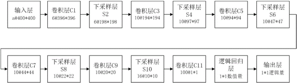
[0056] (2): Construct a thirteen-layer convolutional neural network, such as figure 1 As shown, it includes six convolutional layers, five subsampling layers, one logistic regression layer, and one output layer. Among them, the input is a grayscale image block of 400*400*a, the convolutional layer C1 sets 6 feature maps, the subsampling layer S2 sets 6 feature maps, and the convolution...


PUM


Abstract

The invention discloses a gastrointestinal tumor microscopic hyper-spectral image processing method based on a convolutional neural network, comprising the following steps: reducing and de-noising the spectral dimension of an acquired gastrointestinal tissue hyper-spectral training image; constructing a convolutional neural network structure; and inputting obtained hyper-spectral data principal components (namely, a plurality of 2D gray images, which are equivalent to a plurality of feature maps of an input layer) as input images into the constructed convolutional neural network structure using a batch processing method, and by taking a cross entropy function as a loss function and using an error back propagation algorithm, training the parameters in the convolutional neural network and the parameters of a logistic regression layer according to the average loss function in a training batch until the network converges. According to the invention, the dimension of a hyper-spectral image is reduced using a principal component analysis method, enough spectral information and spatial texture information are retained, the complexity of the algorithm is reduced greatly, and the efficiency of the algorithm is improved.

Description

technical field [0001] The invention relates to the field of medical hyperspectral image processing, in particular to a convolutional neural network-based microscopic hyperspectral image processing method for gastrointestinal tumors. Background technique [0002] Tumor refers to the new organism formed by the proliferation of local tissue cells under the action of various tumorigenic factors, because this new organism is mostly in the form of space-occupying blocky protrusions, also known as neoplasm. Gastrointestinal tumors are common tumors in the digestive tract. Gastrointestinal benign tumors are less likely to recur after resection and are less harmful to the body; gastrointestinal malignant tumors, namely cancer, are the most common malignant tumors with a high incidence worldwide. One, and the incidence rate has increased significantly in recent years. [0003] At present, when performing pathological examination on gastrointestinal tumors, it is necessary to perform...

Claims


Application Information

Patent Timeline
IPC IPC(8): G06T7/00G06T5/00G06N3/08
CPCG06N3/08G06T5/00G06T7/00G06T7/0012G06N3/084G06N3/088G06T5/002G06T2207/10048G06T2207/20081G06T2207/30028G06T2207/30092G06T2207/30096
Inventor 刘治邱清晨肖晓燕曹丽君朱耀文
Owner SHANDONG UNIV
Why Eureka
Try Eureka