
Extraction of information from an image and inclusion thereof in a clinical report

A technology of images, medical images, applied in the field of extracting information from images and including information in clinical reports, which can solve the problems of cost, length and time.

Active Publication Date: 2016-11-30
KONINKLJIJKE PHILIPS NV
View PDF5 Cites 12 Cited by
  • Summary
  • Abstract
  • Description
  • Claims
  • Application Information

AI Technical Summary

Problems solved by technology

Unfortunately, for example, where the reading physician or other medical personnel adds text notes and / or alphanumeric information to the clinical report, the process can be lengthy and time consuming that could otherwise be spent on the patient and / or or read on other images

Method used


Image

  • Extraction of information from an image and inclusion thereof in a clinical report
  • Extraction of information from an image and inclusion thereof in a clinical report
  • Extraction of information from an image and inclusion thereof in a clinical report

Examples


Embodiment Construction

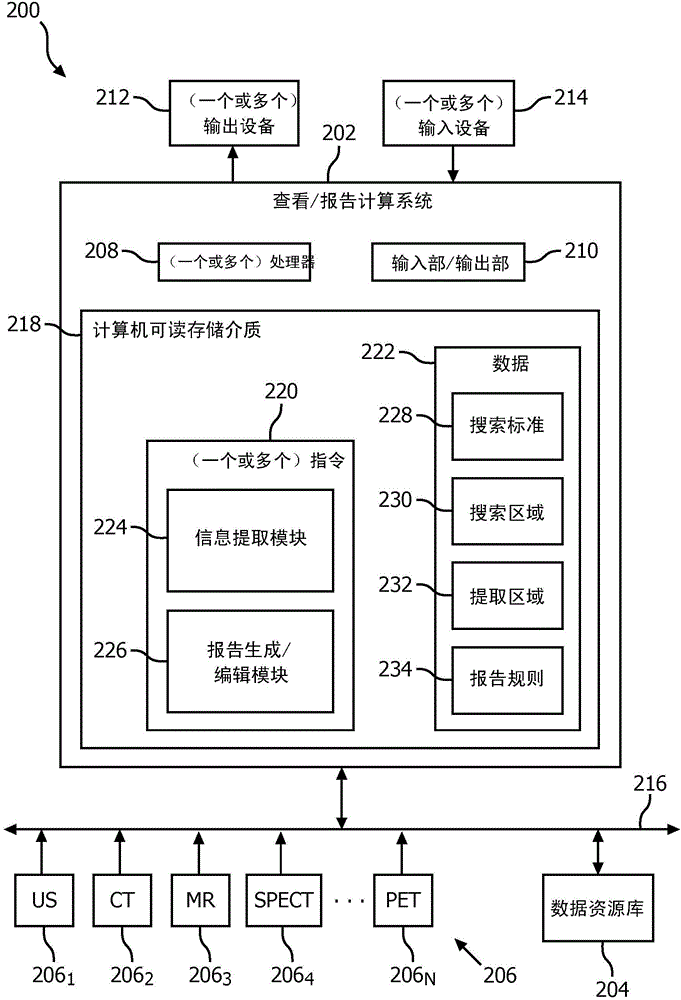
[0022] initial reference figure 2 , schematically illustrates the system 200 . System 200 includes a viewing / reporting computing device 202 coupled to a data repository 204 and N imaging systems 206 , where N is an integer equal to or greater than one. In the illustrated embodiment, viewing / reporting computing device 202 receives images in electronic format from imaging system 206 and / or data repository 204 . The image includes pixels having grayscale values ​​and / or color intensity values ​​and annotation and / or alphanumeric information printed into the image or overlaid on the image.

[0023] N imaging systems 206 include one or more of the following: Ultrasound (US) scanner 206 1 , computed tomography (CT) scanner 206 2 , magnetic resonance (MR) scanner 206 3 , single photon emission computed tomography (SPECT) scanner 206 1 , ..., and a positron emission tomography (PET) scanner 206 N . Data repository 204 includes one or more of the following: Picture Archiving an...


PUM


Abstract

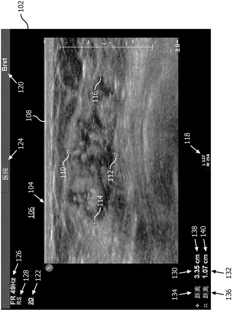
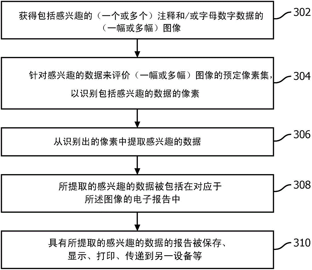
A method includes obtaining, in electronic format, an image (102) including a medical image display region (104) and an information display region (106). The at least one of the medical image display region or the information display region includes graphical indicia representing at least one of an annotation (110, 112, 114, 116) or alphanumeric information (118, 120, 122, 124, 126, 128, 130, 132, 134, 136, 138, 140). The method further includes evaluating pixels of the image to identify pixels including the graphical indicia representing an annotation or alphanumeric information of interest in the image. The method further includes extracting the annotation or alphanumeric information of interest from the identified graphical indicia from the image. The method further includes inserting the extracted annotation or alphanumeric information of interest in an electronically formatted clinical report for the image.

Description

technical field [0001] The following generally relates to extracting information from images and including the extracted information in clinical reports corresponding to the images. Examples of suitable images are images generated by imaging systems such as ultrasound (US) imaging systems, computed tomography (CT) imaging systems, X-ray imaging systems, magnetic resonance (MR) imaging systems, positron A photography emission tomography (PET) imaging system, a single photon emission computed tomography (SPECT) imaging system, and / or other imaging systems. Background technique [0002] figure 1 An example image 102 is shown. Generally, image 102 includes two separate regions 104 and 106 . Area 104 is a medical image display area, and area 106 is an information display area. In the illustrated example, the medical image display area displays annotations ( figure 1 graphics in , but can also include text) of the ultrasound image 108. The annotations include marker pairs 11...

Claims


Application Information

Patent Timeline
Patent Type & Authority Applications(China)
IPC IPC(8): G06K9/32G06K9/20G06K9/00G06F19/00G06V30/10G16H15/00G16H30/40
CPCG16H15/00G16H30/40G06V30/412G06V30/413G06V20/635G06V30/10G06V30/1448G06F40/169
Inventor 邓寅晖林其忠L·博罗茨基李俊杰吴英
Owner KONINKLJIJKE PHILIPS NV
Learn More