

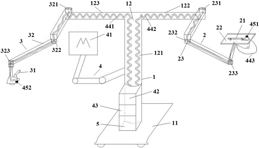
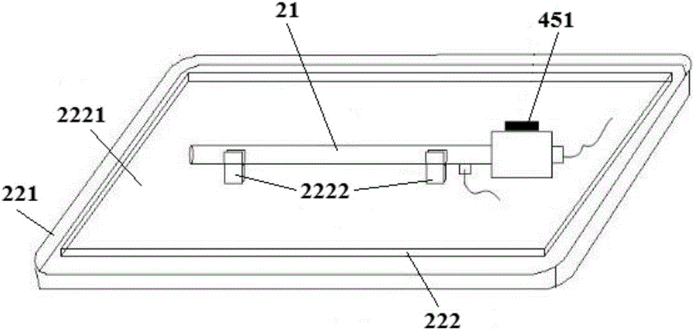
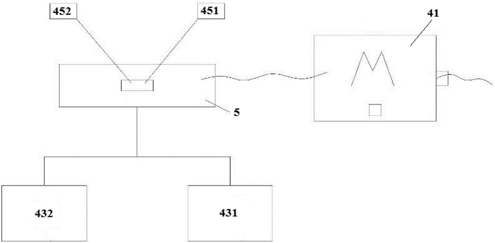
Endoscope and microscope unified body
A technology of microscope and consortium, applied in the fields of surgical microscope, medical science, surgical instrument support, etc., can solve the inconvenience of equipment signal, image display and storage arrangement and editing, endoscope or microscope to achieve satisfactory surgical results, and affect the operation coherence. It can improve the consistency of surgery, shorten the operation time, and facilitate the smooth development of the operation.
- Summary
- Abstract
- Description
- Claims
- Application Information
AI Technical Summary
Problems solved by technology
Method used
Image

Examples
Embodiment Construction
[0041] The following will clearly and completely describe the technical solutions in the embodiments of the present invention with reference to the accompanying drawings in the embodiments of the present invention. Obviously, the described embodiments are only some, not all, embodiments of the present invention. The following description of at least one exemplary embodiment is merely illustrative in nature and in no way taken as limiting the invention, its application or uses. Based on the embodiments of the present invention, all other embodiments obtained by persons of ordinary skill in the art without creative work fall within the protection scope of the present invention.
[0042] Techniques, methods and devices known to those of ordinary skill in the relevant art may not be discussed in detail, but where appropriate, such techniques, methods and devices should be considered part of the Authorized Specification.
[0043]In the description of the present invention, it shoul...
PUM
 Abstract
Description
Claims
Application Information