Fast magnetic resonance heart real-time cine imaging method and fast magnetic resonance heart real-time cine imaging system

An imaging method and imaging system technology, applied in medical imaging, image enhancement, image acquisition, etc., can solve the problems of high scanning time requirements, the acceleration multiple cannot be too large, and the image signal-to-noise ratio reduction.

Active Publication Date: 2017-01-18
SHENZHEN INST OF ADVANCED TECH
View PDF5 Cites 17 Cited by
  • Summary
  • Abstract
  • Description
  • Claims
  • Application Information

AI Technical Summary

Problems solved by technology

[0003] At present, the real-time cine imaging process of magnetic resonance heart needs to collect multiple frames of images in the heart motion cycle, which requires a high scanning time. The commonly used parallel imagin

Method used


Image

  • Fast magnetic resonance heart real-time cine imaging method and fast magnetic resonance heart real-time cine imaging system
  • Fast magnetic resonance heart real-time cine imaging method and fast magnetic resonance heart real-time cine imaging system
  • Fast magnetic resonance heart real-time cine imaging method and fast magnetic resonance heart real-time cine imaging system

Examples


Embodiment 1

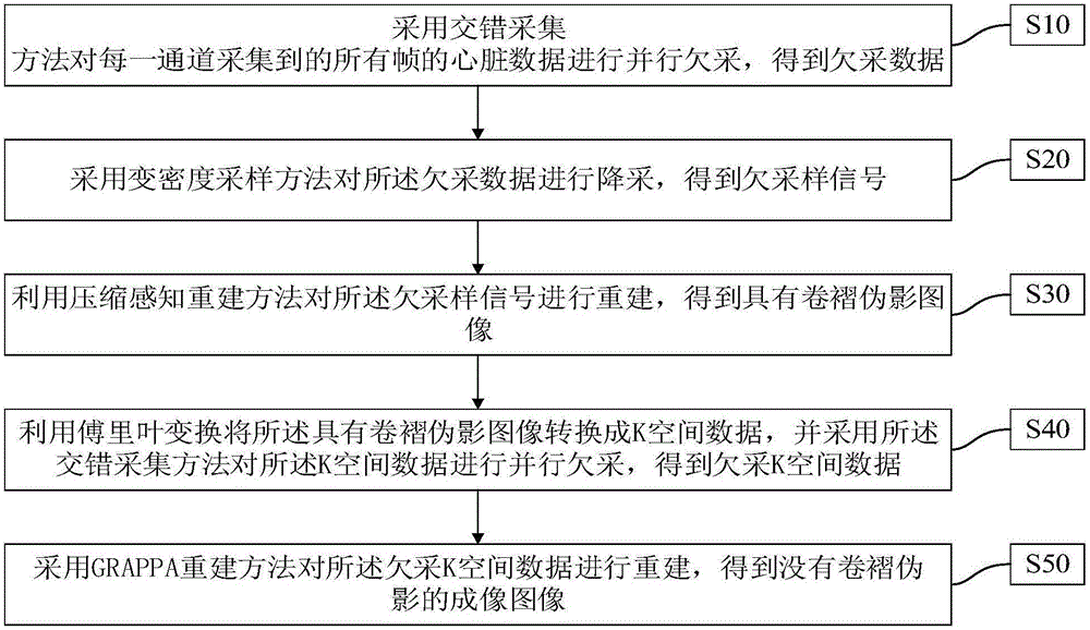
[0066] figure 1 A flow chart showing the fast magnetic resonance cardiac real-time cine imaging method in this embodiment. like figure 1 As shown, the fast magnetic resonance cardiac real-time cine imaging method comprises the following steps:

[0067] S10: Parallel under-acquisition is performed on all frames of heart data collected by each channel by using an interleaved acquisition method to obtain under-acquisition data. Undersampling refers to undersampling in one dimension (such as phase encoding direction) or multiple dimensions. Specifically, the interleaved acquisition method includes the following steps: preset the sampling rate of each frame of data as R 并行 , the frame number of data acquisition is N phase , the number of phase codes is N pe ; For each frame of data, the frequency encoding direction is fully sampled, and the phase encoding direction is collected every R 并行 -1 to acquire a line; and the nRth 并行 +R frame data is collected from the Rth line unti...

Embodiment 2

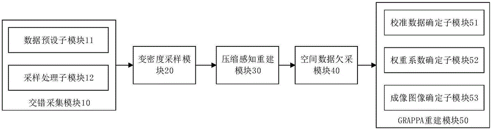
[0110] figure 2 A functional block diagram of the fast magnetic resonance cardiac real-time cine imaging system in this embodiment is shown. like figure 2 As shown, the fast magnetic resonance cardiac real-time cine imaging system includes an interleaved acquisition module 10 , a variable density sampling module 20 , a compressed sensing reconstruction module 30 , a spatial data underacquisition module 40 and a GRAPPA reconstruction module 50 .

[0111] The interlaced acquisition module 10 is configured to perform parallel under-acquisition on the heart data of all frames collected by each channel by adopting an interleaved acquisition method to obtain under-acquisition data. Undersampling refers to undersampling in one dimension (such as phase encoding direction) or multiple dimensions. Specifically, the interleaved acquisition module 10 includes a data preset submodule 11 and a sampling processing submodule 12 .

[0112] The data preset sub-module 11 is used to preset t...


PUM


Abstract

The invention discloses a fast magnetic resonance heart real-time cine imaging method and a fast magnetic resonance heart real-time cine imaging system. The fast magnetic resonance heart real-time cine imaging method comprises the following steps: under-sampling all frames of heart data collected by each channel in parallel through use of a staggered collection method to get under-sampled data; down-sampling the under-sampled data through use of a variable-density sampling method to get an under-sampled signal; reconstructing the under-sampled signal through use of a compressed sensing reconstruction method to get an image with wraparound artifacts; converting the image with wraparound artifacts into K-space data through Fourier transform, and under-sampling the K-space data in parallel through use of the staggered collection method to get under-sampled K-space data; and reconstructing the under-sampled K-space data through use of a GRAPPA reconstruction method to get an imaging image without wraparound artifacts. According to the fast magnetic resonance heart real-time cine imaging method and the fast magnetic resonance heart real-time cine imaging system, wraparound artifacts can be removed without additionally collecting K-space data, an image of good quality can be obtained at higher acceleration multiple, and there is no need for a subject to hold his/her breath during scanning.

Description

technical field [0001] The invention relates to the technical field of magnetic resonance imaging, in particular to a fast magnetic resonance cardiac real-time movie imaging method and system. Background technique [0002] Magnetic resonance cardiac real-time cine imaging is an imaging mechanism that obtains a series of images of cardiac motion at a high frame rate. It is often used clinically to detect cardiac function. During the scan, the subject needs to hold his breath to obtain multiple cardiac motion cycles of the subject. The complete K-space data within. However, if the subject suffers from heart disease or the subject is a child, it is often difficult to cooperate with repeated breath-holding requirements, and the scanning time cannot be too long. Therefore, it is necessary to reduce the amount of data collected in each frame as much as possible on the premise that the imaging quality is clinically acceptable, and increase the sampling speed, thereby reducing the ...

Claims


Application Information

Patent Timeline
IPC IPC(8): G06T3/20G06T1/00A61B5/055G06T5/00
CPCA61B5/055A61B2576/023G06T1/0007G06T3/20G06T5/002G06T2207/10088G06T2207/20056G06T2207/30048
Inventor 梁栋刘元元朱燕杰刘新郑海荣
Owner SHENZHEN INST OF ADVANCED TECH
Why Eureka
Try Eureka