A helical scanning method and device

A technology of helical scanning and actual scanning, applied in the field of medical devices, can solve problems such as unsatisfactory images and insufficient scanning area, and achieve the effects of ensuring data continuity, reducing radiation, and saving scanning doses

Active Publication Date: 2019-11-12
NEUSOFT MEDICAL SYST CO LTD
View PDF5 Cites 0 Cited by
  • Summary
  • Abstract
  • Description
  • Claims
  • Application Information

AI Technical Summary

Problems solved by technology

[0004] After a helical scan, if it is found that the scan area is not long enough, the scan is too short (not enough), or if a certain part of the image is found to be unsatisfactory, the doctor needs to re-plan a scan, and continue the scan at the end of the previous scan. or rescan undesired areas

Method used


Image

  • A helical scanning method and device
  • A helical scanning method and device
  • A helical scanning method and device

Examples


Embodiment Construction

[0048] In order to make the above objects, features and advantages of the present invention more obvious and understandable, the main technical idea of ​​the present invention will be explained below.

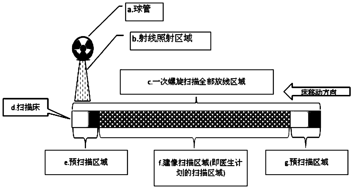
[0049] In the prior art, the helical scanning device regards each helical scan as an independent helical scan. When the helical scanning device receives the helical scan instruction, it will directly perform the helical scan according to the scanning parameters, and obtain The raw data can complete the imaging. When building an image, it is necessary to rely on the scan data of the adjacent front and rear positions of a position to complete the image building of the position. see figure 1 , figure 1 The helical scanning actual scene example figure provided for the present invention, in figure 1 Among them, a represents the tube, b represents the area irradiated by radiation, d represents the scanning bed, and c represents the entire line-emitting area of ​​a helical scan, in...


PUM


Abstract

The invention provides a spiral scanning method and device. The two schemes are consistent in technological thought. The method comprises the following steps: determining an overlap area between the scanning area of current spiral scanning and the scanning area of previous spiral scanning; starting current spiral scanning, controlling a bulb tube not to emit rays in the overlap area but in the non-overlap area of current spiral scanning, and scanning to obtain raw data of current spiral scanning; and performing data fusion of the raw data of current spiral scanning and the raw data obtained by previous spiral scanning. Through the technical scheme provided by the invention, in a scene of repeated spiral scanning, the scanning of a certain area can be saved, and the radiation of the scanned person is reduced to a certain degree; and more importantly, the data continuity is guaranteed through a way of data fusion, and complete data is provided for the imaging of current spiral scanning.

Description

technical field [0001] The invention relates to the technical field of medical devices, in particular to a computer tomography helical scanning method and device. Background technique [0002] In the process of computer tomography (Computed Tomography, CT), the X-ray tube rotates continuously around the gantry for exposure, and the examination bed moves synchronously and uniformly at the same time as the exposure, and the detector collects data. Because the scanning trajectory is helical, it is called helical scanning, also known as volume or volume scanning. [0003] In a clinical environment, when doctors use helical scanning, they often encounter the following situations: [0004] After a helical scan, if it is found that the scan area is not long enough, the scan is too short (not enough), or if a certain part of the image is found to be unsatisfactory, the doctor needs to re-plan a scan, and continue the scan at the end of the previous scan. Or rescan undesired areas....

Claims


Application Information

Patent Timeline
Patent Type & Authority Patents(China)
IPC IPC(8): A61B6/03
CPCA61B6/027A61B6/032A61B6/52A61B6/54A61B6/542
Inventor 李伟
Owner NEUSOFT MEDICAL SYST CO LTD
Why Eureka
Try Eureka