Radiological image detection apparatus, radiographic apparatus and radiographic system

a detection apparatus and radiographic technology, applied in the direction of diaphragms for radiation diagnostics, tomography, nuclear engineering, etc., can solve the problems of reducing the absorption ability of x-rays, difficult to obtain the inability to obtain sufficient contrast of images, so as to reduce the scattering

Inactive Publication Date: 2012-06-28
FUJIFILM CORP
View PDF1 Cites 66 Cited by
  • Summary
  • Abstract
  • Description
  • Claims
  • Application Information

AI Technical Summary

Benefits of technology

[0021]According to the invention, it is possible to reduce the scatte

Problems solved by technology

However, the X-ray absorption ability is reduced in case of the material consisting of the element having the smaller atomic number.
Accordingly, for the soft biological tissue or soft material, it is not possible to obtain the sufficient contrast of an image for the X-ray absorption contrast image.
Thus, since a difference of the X-ray absorption amounts thereof is small, it is difficult to obtain the sufficient contrast of the images.
When manufacturing the first or second grating, which is used for the X-ray phase contrast imaging, in the high aspect ratio capable of securing the scattering removal function, there are many difficulties such as difficulty in forming strips in a high aspect ratio by an etching and the like, non-uniformity of a substrate thickness, in-plane uniformity

Method used


Image

  • Radiological image detection apparatus, radiographic apparatus and radiographic system
  • Radiological image detection apparatus, radiographic apparatus and radiographic system
  • Radiological image detection apparatus, radiographic apparatus and radiographic system

Examples


Embodiment Construction

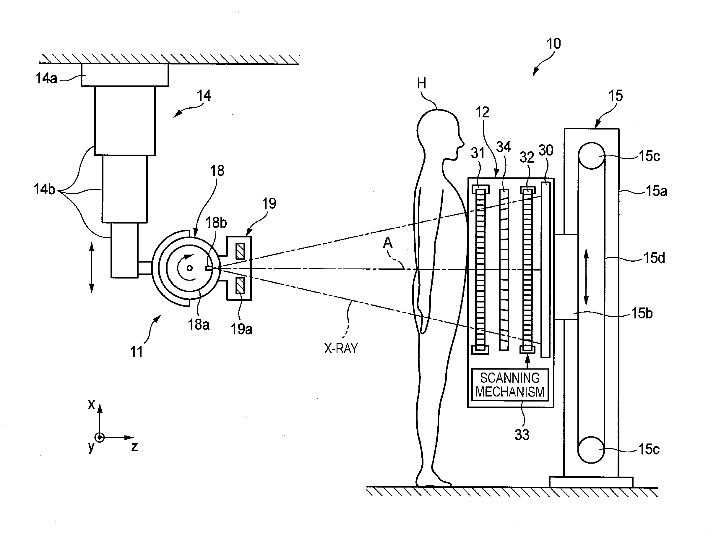
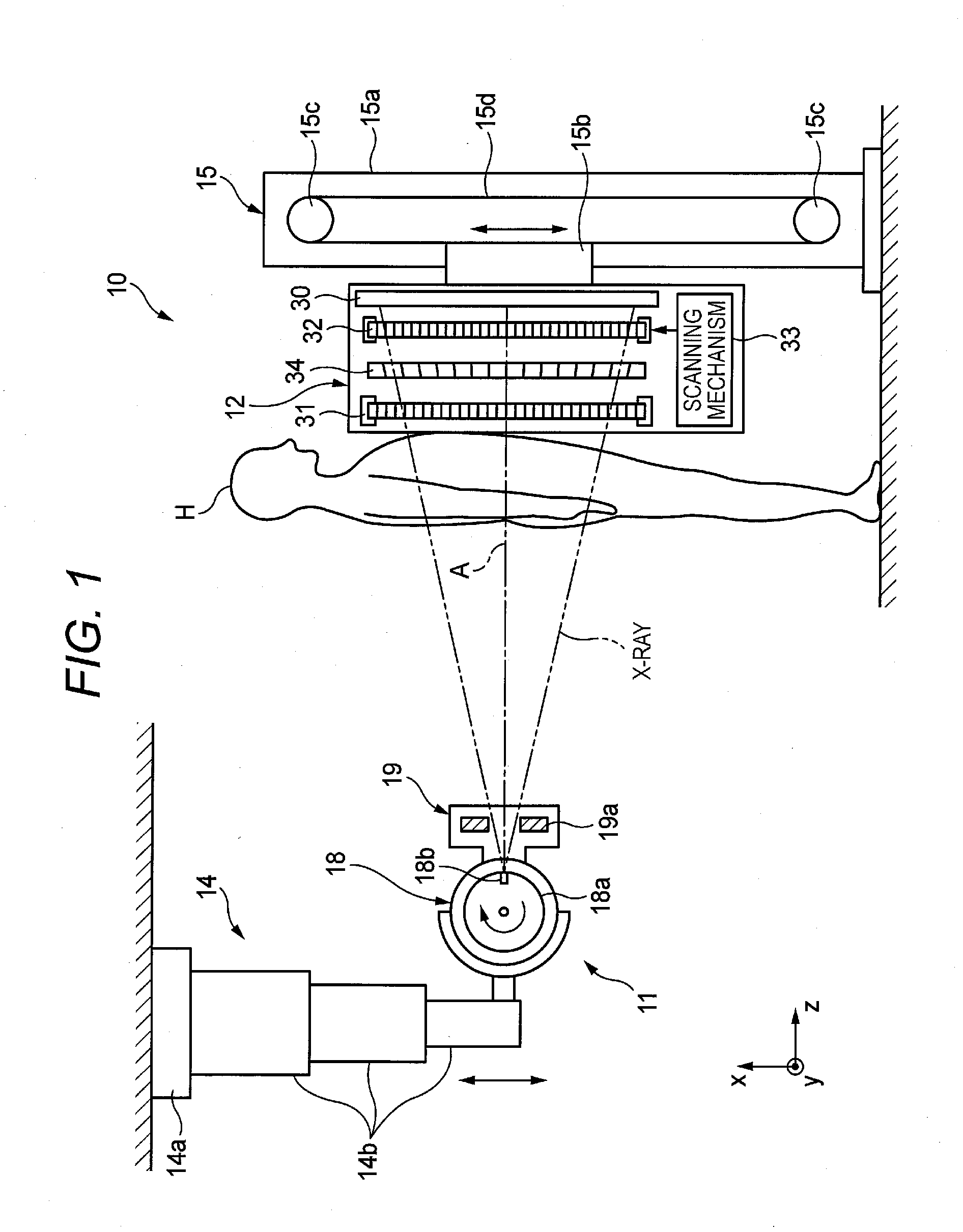
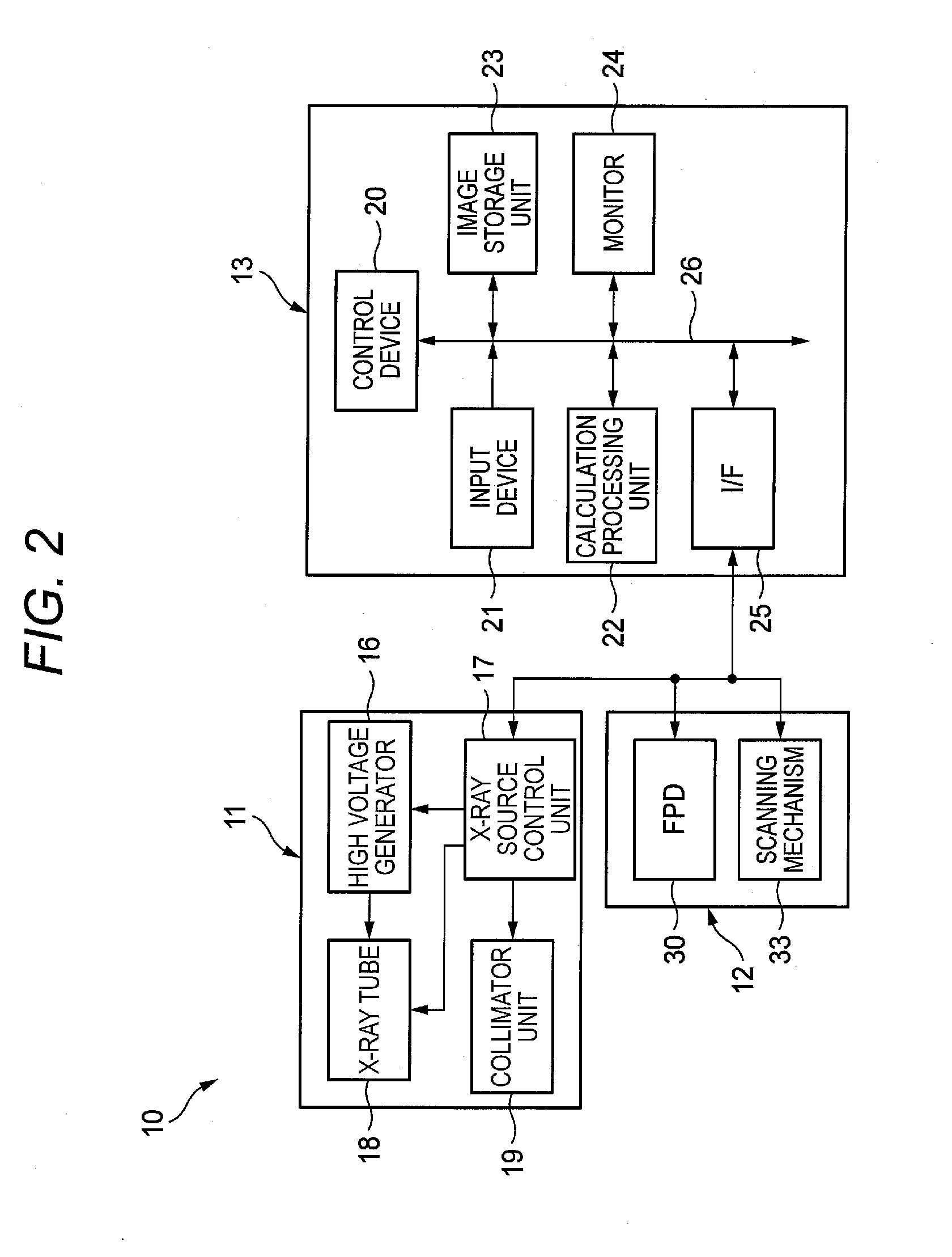
[0061]FIG. 1 shows an example of a configuration of a radiographic system for illustrating an illustrative embodiment of the invention and FIG. 2 shows a control block diagram of the radiographic system of FIG. 1.

[0062]In the meantime, the same configurations as those already described are indicated with the same reference numerals and the descriptions thereof are omitted. The differences from the configurations already described will be described.

[0063]An X-ray imaging system 10 is an X-ray diagnosis apparatus that performs an imaging while a subject (patient) H stands, and includes an X-ray source 11 that radiates the subject H, an imaging unit 12 functioning as a radiological image detection apparatus that is opposed to the X-ray source 11 with the subject H being interposed between the X-ray source 11 and the imaging unit, detects the X-ray having penetrated the subject H from the X-ray source 11 and thus generates image data and a console 13 (refer to FIG. 2) that controls an e...


PUM


Abstract

A radiological image detection apparatus includes a first grating unit, a grating pattern unit, a radiological image detector, and an anti-scatter grating. The grating pattern unit has a period that substantially coincides with a pattern period of a radiological image formed by radiation having passed through the first grating unit. The radiological image detector detects the radiological image masked by the grating pattern unit. The anti-scatter grating is arranged on a path of the radiation incident onto the radiological image detector and removes scattered radiation. A smoothing process is performed for at least one of a surface and a backside of the anti-scatter grating intersecting with a traveling direction of the radiation.

Description

CROSS-REFERENCE TO RELATED APPLICATIONS[0001]This application claims the benefit of Japanese Patent Application No. 2010-286767 (filed on Dec. 22, 2010), the entire contents of which are hereby incorporated by reference.BACKGROUND[0002]1. Technical Field[0003]The invention relates to a radiological image detection apparatus, a radiographic apparatus and a radiographic system capable of enabling a phase contrast imaging of a subject by using radiation such as X-ray.[0004]2. Description of Related Art[0005]Since X-ray attenuates depending on an atomic number of an element configuring a material and a density and a thickness of the material, it is used as a probe for seeing through an inside of a subject. An imaging using the X-ray is widely spread in fields of medical diagnosis, nondestructive inspection and the like.[0006]In a general X-ray imaging system, a subject is arranged between an X-ray source that irradiates the X-ray and an X-ray image detector that detects the X-ray, and a...

Claims


Application Information

Patent Timeline
IPC IPC(8): G21K1/10
CPCA61B5/45A61B6/06A61B6/4035A61B6/4233A61B6/4021A61B6/4452A61B6/4464A61B6/484A61B6/502A61B6/4291
Inventor TADA, TAKUJI
Owner FUJIFILM CORP
Why Eureka
Try Eureka