Imaging biomarker for guiding percutaneous vertebroplasty and application of imaging biomarker for guiding percutaneous vertebroplasty

A biomarker, vertebroplasty technology, which is applied in the fields of radiological diagnosis instruments, medical imaging, surgery, etc., can solve the problems of difficulty in quantitative analysis and evaluation of surgical parameters, difficulty in obtaining clear and intuitive images, etc. Reduce the possibility of serious complications, reduce the effect of exposure

Inactive Publication Date: 2017-02-22
GUANGXI UNIVERSITY OF TECHNOLOGY
View PDF2 Cites 3 Cited by
  • Summary
  • Abstract
  • Description
  • Claims
  • Application Information

AI Technical Summary

Problems solved by technology

Due to the particularity of vertebral body anatomy, it is difficult to obtain clear and intuitive images using conventional medical images
Although orthopedic surgeons can rely on

Method used


Image

  • Imaging biomarker for guiding percutaneous vertebroplasty and application of imaging biomarker for guiding percutaneous vertebroplasty

Examples


Embodiment Construction

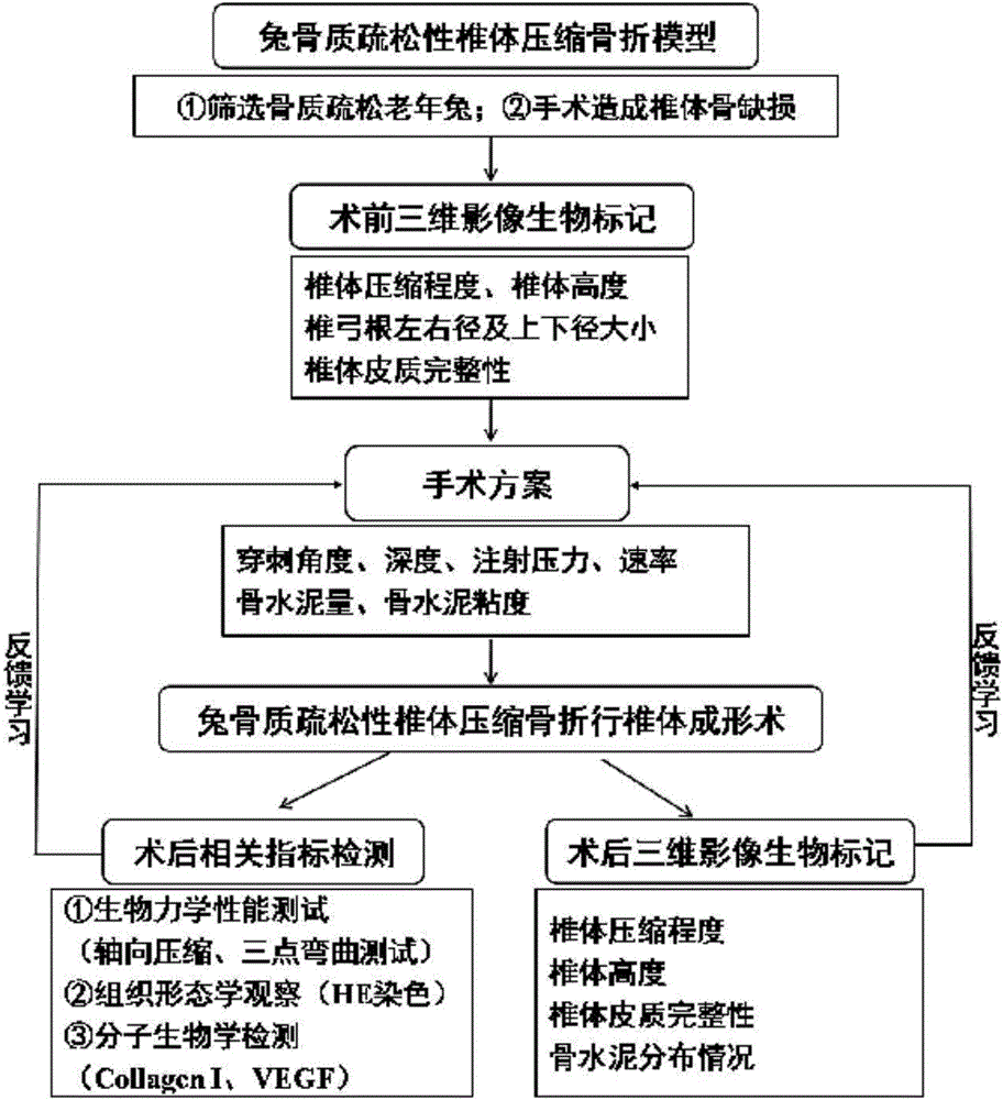
[0012] The present invention is used to guide the imaging biomarkers of percutaneous vertebroplasty, the imaging biomarkers include preoperative imaging biomarkers and postoperative imaging biomarkers, and the preoperative imaging biomarkers are respectively: vertebral body compression degree , vertebral body height, pedicle left and right diameters and upper and lower diameters, and vertebral cortex integrity; postoperative imaging biomarkers were: vertebral body compression degree, vertebral body height, vertebral body cortex integrity, and bone cement distribution.

[0013] Imaging biomarkers are obtained through the following methods: use CT machine to obtain preoperative and postoperative CT images, further use open source software (such as the 3D Viewer plug-in expansion package on the ImageJ platform) to complete 3D reconstruction of CT images, and then perform 3D reconstruction on CT images. In the process, the optical property model including the X-ray tube response, t...


PUM


Abstract

The invention relates to a marker in medicine and application of the marker, in particular to an imaging biomarker for guiding percutaneous vertebroplasty and application of the imaging biomarker for guiding percutaneous vertebroplasty. The imaging biomarker comprises a preoperative imaging biomarker and a postoperative imaging biomarker, wherein the preoperative imaging biomarker involves vertebral compression degree, a vertebral height, a vertebral pedicle transversal diameter, a vertebral pedicle vertical diameter and vertebral cortex integrity, and the postoperative imaging biomarker involves vertebral compression degree, a vertebral height, vertebral cortex integrity and bone cement distribution conditions. The imaging biomarker provides quantitative bases for operation plans and lays a firm foundation for precise standardized vertebroplasty plans.

Description

technical field [0001] The invention relates to a marker in medicine and its use, in particular to quantitative or semi-quantitative parameters based on three-dimensional reconstruction image analysis as image biomarkers. Background technique [0002] Percutaneous vertebroplasty (PVP) for thoracolumbar fractures fixes microfractures of the vertebrae by injecting bone cement into the vertebrae with recent compression fractures and kills the sensory nerve endings in the vertebral body through high fever (average of about 75 degrees in the center), So as to achieve the purpose of relieving the patient's pain and restoring function. Internationally, Gakibert used PVP clinically for the first time in 1987, and it has been used in my country since 1999, because of its relatively low invasiveness, low risk, low cost, and generally the advantages of immediate and long-term relief of patient pain, it has become a A common treatment in orthopedics, it is the first choice to treat seve...

Claims


Application Information

Patent Timeline
IPC IPC(8): A61B34/10A61B6/03
CPCA61B6/032A61B6/5211A61B2576/02
Inventor 刘洪李龙蓝赠美吕亚萍徐奕奕李厚君王日风
Owner GUANGXI UNIVERSITY OF TECHNOLOGY
Why Eureka
Try Eureka