

Method for extracting liver region in ultrasound image
An ultrasound image and a technology in the image, which is applied in the field of extracting the liver area in the ultrasound image, can solve the problems of large noise in the liver area and difficulty in extracting completeness, and achieve a good extraction effect
- Summary
- Abstract
- Description
- Claims
- Application Information
AI Technical Summary
Problems solved by technology
Method used
Image

Examples
Embodiment Construction
[0034] The present invention will be described in further detail below in conjunction with the accompanying drawings and embodiments. It should be understood that the specific embodiments described here are only used to explain the present invention, not to limit the present invention.
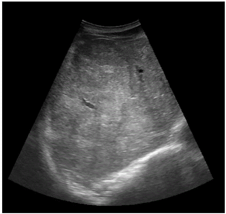
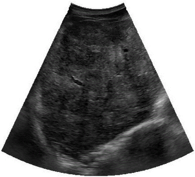
[0035] Figure 1 to Figure 7 It schematically shows a method for extracting the liver region in the ultrasound image according to the disclosure of the present invention.
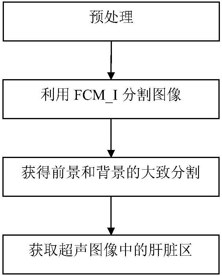
[0036] Such as figure 1As shown, a method for extracting liver regions in ultrasound images disclosed in the present invention uses the FCM algorithm and uses prior knowledge to control the connection of classified regions. However, FCM is only a clustering algorithm, so it needs to cooperate with other pre-processing and post-processing to complete the task of extracting liver regions. The following is the liver region extraction scheme proposed by the present invention.
[0037] Step 1: Preprocessing, specifically, de...
PUM
 Abstract
Description
Claims
Application Information