
Medical image analysis method and device

A medical image and analysis method technology, applied in the field of medical image analysis methods and devices, can solve the problems of false positives, insufficient features to describe and distinguish lesions and normal areas, etc., to improve accuracy, overcome insufficient feature extraction, Accurate and quick results

Inactive Publication Date: 2017-03-22
朱育盼
View PDF5 Cites 7 Cited by
  • Summary
  • Abstract
  • Description
  • Claims
  • Application Information

AI Technical Summary

Problems solved by technology

[0003] However, the application of existing technologies in gastrointestinal endoscopy will produce a large number of false positives, mainly because the models used by such methods usually only contain a hidden layer for extracting features, and the extracted features are often not enough to describe and distinguish lesions point and normal area

Method used


Image

  • Medical image analysis method and device
  • Medical image analysis method and device

Examples


Embodiment Construction

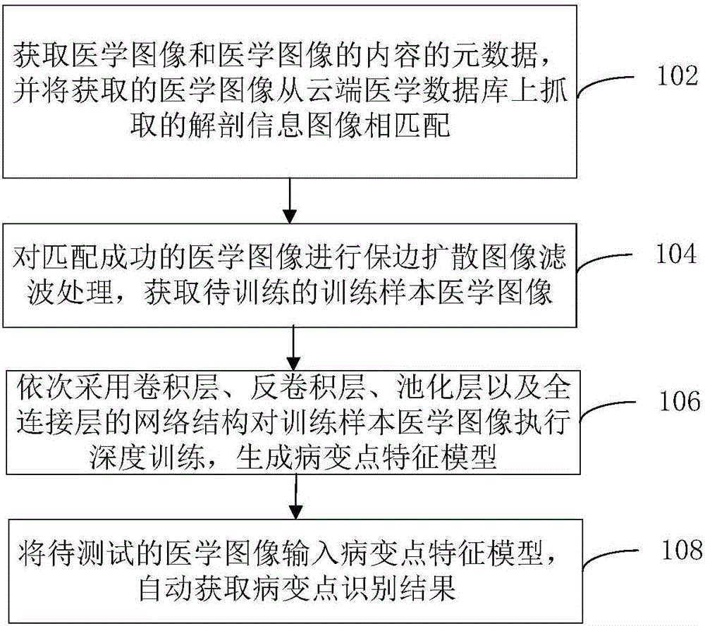
[0018] In order to make the purpose, technical solution and advantages of the present invention clearer, the medical image analysis method and device of the present invention will be further described in detail below with reference to the accompanying drawings and embodiments. It should be understood that the specific embodiments described here are only used to explain the present invention, and are not intended to limit the present invention.

[0019] It should be noted that the disadvantages of the prior art have been explained in the background art. The neural network used in the deep learning solution adopted in this technical solution has the characteristics of extracting high-level features of objects. Since the high-level feature information is a linear and nonlinear transformation of the underlying feature information, the deep neural network can extract the essential features that can describe the object to be described better than the shallow network, thereby improvi...


PUM


Abstract

The present invention relates to a medical image analysis method and device. The method includes the following steps that: medical images and the metadata of the content of the medical images are acquired, and the acquired medical images are matched with anatomical information images captured from a cloud medical database; edge-preserving diffusion image filtering processing is performed on medical images which are successfully matched with the anatomical information images, so that training sample medical images to be trained are obtained; network structures of a convolution layer, a de-convolution layer, a pooling layer and a whole-connection layer are sequentially adopted to perform deep training on the training sample medical images, so that a lesion point feature model is generated; and a medical image to be tested is inputted into the lesion point feature model, and a lesion point recognition result is automatically obtained. With the above method adopted, the accuracy of an algorithm can be greatly improved, false negatives can be decreased, a lesion point can be obtained accurately and rapidly, and further analysis operation can be performed on the lesion point; and a deep network is adopted, so that the problem of insufficiency of shallow-layer network feature extraction can be solved.

Description

technical field [0001] The present invention relates to the technical field of image processing, in particular to a medical image analysis method and device. Background technique [0002] Using algorithms to automatically identify lesions or potential lesions such as fatty liver and intestinal polyps from medical images is a problem that people have been trying to solve for many years. The traditional computer automatic recognition algorithm is to convert the original image input, that is, the pixel value into manually defined features, such as SIFT, HOG features, etc.; and then put these transformed features into a pre-trained shallow detector. Detection, the detection process is generally, slide a detection window with a preset size on the original picture, if the detection score calculated at a certain position is higher than a certain preset threshold, it is considered that this position exists. Lesion of interest or potential lesion. [0003] However, the application ...

Claims


Application Information

Patent Timeline
IPC IPC(8): G06T7/00
CPCG06T7/0012G06T2207/20081
Inventor 朱育盼
Owner 朱育盼
Learn More