Visualizing different types of airway wall abnormalities

An airway wall and visualization technology, which is applied in the directions of radiological diagnosis instruments, image enhancement, instruments, etc., can solve the problems of airway wall thickness and response to treatment, etc.

Active Publication Date: 2017-04-05
SIEMENS HEALTHCARE GMBH
View PDF3 Cites 74 Cited by
  • Summary
  • Abstract
  • Description
  • Claims
  • Application Information

AI Technical Summary

Problems solved by technology

[0006] However, airway walls may be thicker due to inflammatory disease processes or due to other processes such as scarring
Also, inflammatory causes of thickening may be potentially treatable (such as with anti-inflammatory agents), but scar-type processes are less likely to respond to treatment

Method used


Image

  • Visualizing different types of airway wall abnormalities
  • Visualizing different types of airway wall abnormalities
  • Visualizing different types of airway wall abnormalities

Examples


Embodiment Construction

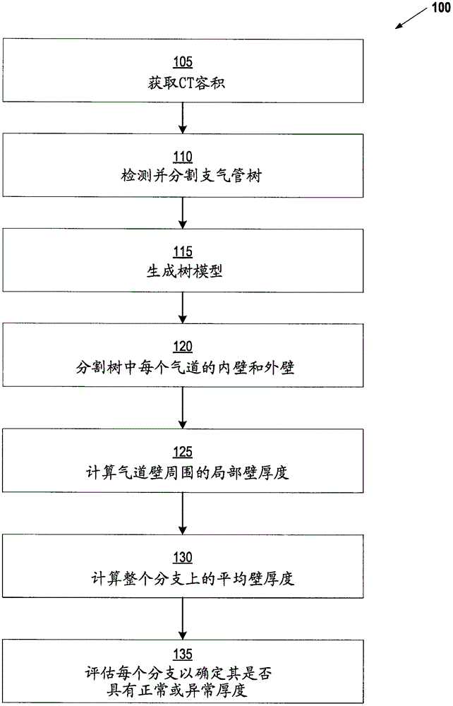
[0022] The following disclosure describes the invention in terms of several embodiments directed to methods, systems and devices related to visualizing different types of airway wall abnormalities. The techniques described herein can be used to automatically calculate and display the location of airways that are: (i) thickened but not inflamed; (ii) thickened and inflamed; (iii) inflamed but not thickened; or (iv) )normal. Any detected abnormalities can be presented in an interactive display that allows the physician to quickly identify these different types of abnormalities. Such a display gives physicians the ability to distinguish thickened walls that are inflamed and potentially treatable with anti-inflammatory drugs from non-inflamed thickened walls that have a less favorable prognosis.

[0023] figure 1 A method 100 for detecting thickened bronchial walls is presented according to some embodiments. The method begins at step 105 by acquiring a CT volume of the patient'...


PUM


Abstract

The invention relates to visualizing different types of airway wall abnormalities. A method for visualizing airway wall abnormalities includes acquiring Dual Energy Computed Tomography (DECT) imaging data comprising one or more image volumes representative of a bronchial tree. An iodine map is derived using the DECT imaging data and the bronchial tree is segmented from the image volume(s). A tree model representative of the bronchial tree is generated. Then, for each branch, this tree model is used to determine an indicator of normal or abnormal thickness. Locations corresponding to bronchial walls in the bronchial tree using the tree model are identified. Next, for each branch, the locations corresponding to bronchial walls in the bronchial tree and the iodine map are used to determine an indicator of normal or abnormal inflammation. A visualization of the bronchial tree may be presented with visual indicators at each of the locations corresponding to bronchial walls indicating whether a bronchial wall is thickened and/or inflamed.

Description

[0001] Cross References to Related Applications [0002] This application claims the benefit of US Provisional Application Serial No. 62 / 221,700, filed September 22, 2015, which is incorporated herein by reference in its entirety. technical field [0003] The present invention generally relates to methods, systems and devices for visualizing different types of airway wall abnormalities. The techniques described herein can be applied, for example, to the clinical diagnosis of lung disease. Background technique [0004] Lung diseases such as bronchiectasis, asthma, cystic fibrosis, and chronic obstructive pulmonary disease (COPD) are characterized by abnormalities in airway dimensions, including airway wall thickness and lumen size (inner airway )). Computed tomography (CT) has become one of the primary means of delineating and detecting these abnormalities, as the availability of high-resolution near-homotropic data has made it possible to assess the airway at angles obliqu...

Claims


Application Information

Patent Timeline
Patent Type & Authority Applications(China)
IPC IPC(8): A61B6/03G06T7/00
CPCA61B6/032A61B6/48A61B6/5205A61B6/5217G06T7/0012G06T2207/30061G06T2207/10081G06T11/008G06T2207/30101G06T2211/408A61B6/482A61B6/50A61B6/5223G06T7/11
Inventor A·基拉利C·诺瓦克B·奥德里
Owner SIEMENS HEALTHCARE GMBH
Why Eureka
Try Eureka