SD-OCT retina image CNV segmentation method based on concavity and convexity

A retinal and concavo-convex technology, applied in the field of CNV automatic segmentation of retinal images based on frequency-domain optical coherence tomography, to overcome blurred or even missing lesion boundaries and the effect of overcoming the influence of CNV segmentation

Active Publication Date: 2017-04-26
NANJING UNIV OF SCI & TECH
View PDF3 Cites 24 Cited by
  • Summary
  • Abstract
  • Description
  • Claims
  • Application Information

AI Technical Summary

Problems solved by technology

[0003] At present, there are only automatic CNV segmentation methods based on OCT angiography images, and there is no automatic CNV segmentation method based on frequency-domain OCT (SD-OCT), because CNVs in SD-OCT images cannot be distinguished by color differences like OCT angiography images. easier to distinguish

Method used


Image

  • SD-OCT retina image CNV segmentation method based on concavity and convexity
  • SD-OCT retina image CNV segmentation method based on concavity and convexity
  • SD-OCT retina image CNV segmentation method based on concavity and convexity

Examples


Embodiment

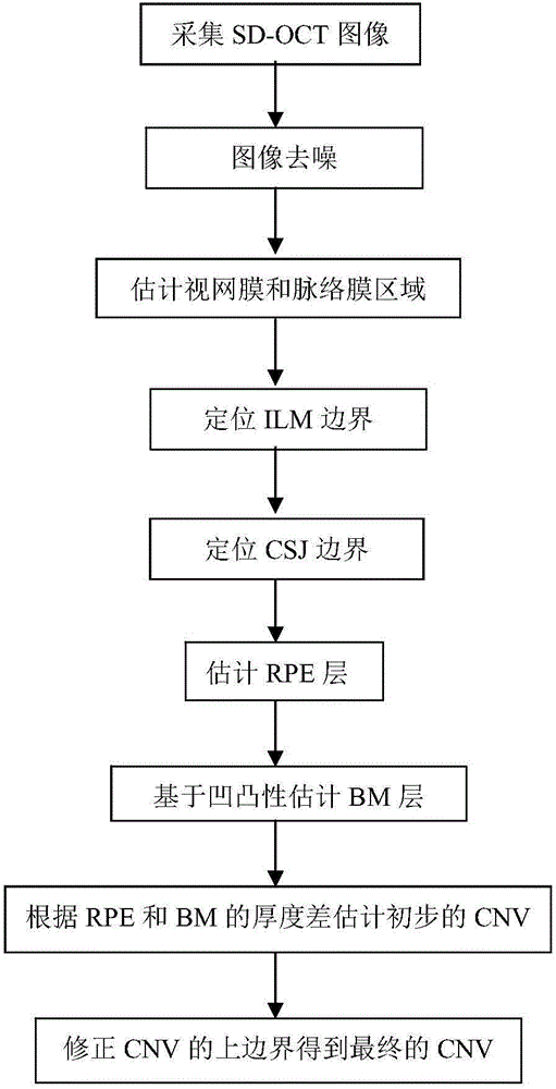
[0086] The invention of the system uses SD-OCT retinal images as input, and uses image processing means to automatically segment the CNV in the input image.

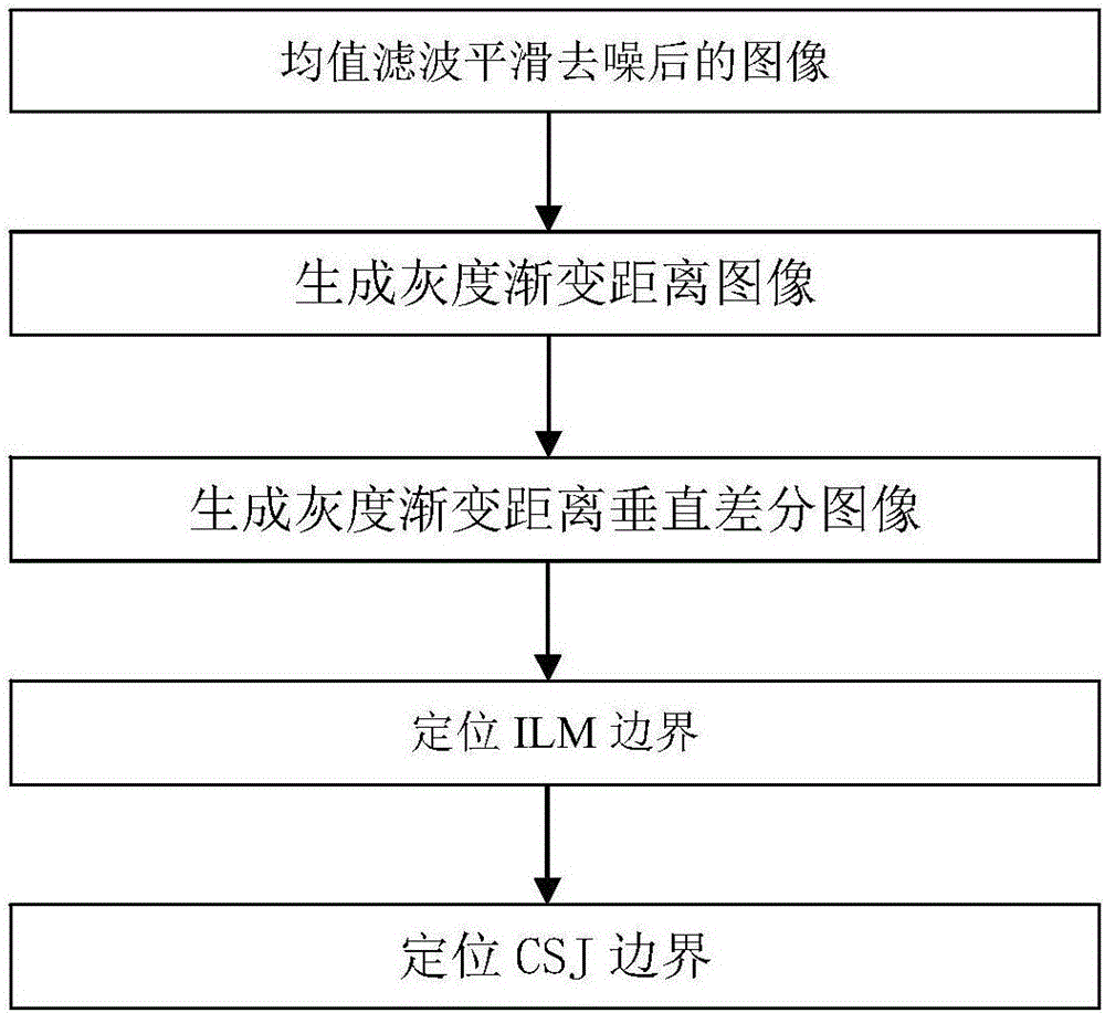
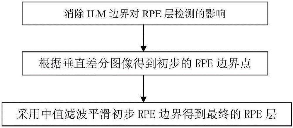
[0087] The flow of this embodiment is as follows figure 1 As shown, the size of the three-dimensional SD-OCT retinal image collected by the OCT imaging device is 1024×512×128, corresponding to the retinal area of ​​2mm×6mm×6mm, Figure 7 A frame of original SD-OCT retinal image is given, and several main related tissue structures of the retina are marked in the figure (ILM: inner limiting membrane, RPE: retinal pigment epithelium, BM: Bruch's membrane, CNV: choroidal neogenesis Vascular, CSI: chorioscleral border). Figure 8 is the smoothing result of bilateral filtering, Figure 9 The retinal and choroidal areas estimated based on the reflectance characteristics can be easily obtained by a global threshold because the reflective rates of the retinal and choroidal areas are significantly higher than those of other area...


PUM


Abstract

The invention discloses a spectral-domain optical coherence tomography (SD-OCT) retina image choroidal neovascularization (CNV) segmentation method based on concavity and convexity, and belongs to the technical field of image processing. The method comprises the following steps: first, estimating the retina and choroid area of an input SD-OCT image, and positioning the internal limiting membrane (ILM) and the choroid-sclera junction (CSJ); then, estimating the retina pigment epithelium (RPE) layer according to the gradual change feature of the reflectivity of the retina image, and estimating the Bruch's membrane (BM) layer based on the concavity and convexity of the RPE layer; and finally, estimating a preliminary CNV region according to the thickness difference between the RPE layer and the BM layer, and correcting the upper border of CNV to get a final CNV segmentation result. The experimental results show that the algorithm proposed in the invention can be used to segment CNV robustly and precisely, and is of great significance to facilitating subsequent CNV quantitative analysis and improving the work efficiency of doctors.

Description

technical field [0001] The invention relates to an automatic CNV segmentation method, in particular to an automatic CNV segmentation method based on frequency-domain optical coherence tomography (SD-OCT) retinal images. Background technique [0002] Choroidal neovascularization (CNV) is a major manifestation of advanced age-related macular degeneration (AMD), which can lead to subretinal hemorrhage, subretinal fluid exudation and other lesions that affect vision. Traditional CNV lesion measurement is mainly based on two-dimensional imaging techniques such as fluorescein angiography and indigo green angiography. Optical coherence (OCT) imaging technology can effectively obtain three-dimensional images of the fundus. Based on OCT angiography images, three-dimensional parameters such as the volume of CNV can be measured. Thus, CNV can be analyzed more effectively. [0003] At present, there are only automatic CNV segmentation methods based on OCT angiography images, and there ...

Claims


Application Information

Patent Timeline
Patent Type & Authority Applications(China)
IPC IPC(8): G06T7/12
Inventor 陈强俞晨琛李鸣超李苹
Owner NANJING UNIV OF SCI & TECH
Why Eureka
Try Eureka