
Brain glioma molecular marker nondestructive prediction method and prediction system based on radiomics
A technology of molecular markers and radiomics, applied in image analysis, image enhancement, image data processing, etc., can solve problems such as lack of research on analysis and understanding
- Summary
- Abstract
- Description
- Claims
- Application Information
AI Technical Summary
Problems solved by technology
Method used
Image

Examples
Embodiment Construction
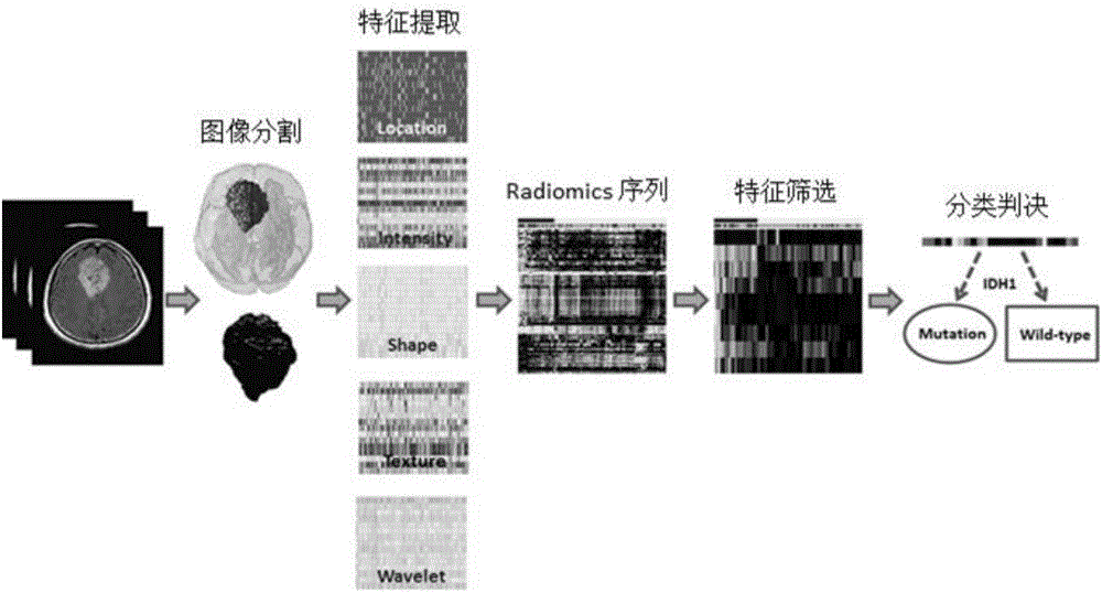
[0060] The following are the specific implementation steps of the entire algorithm:
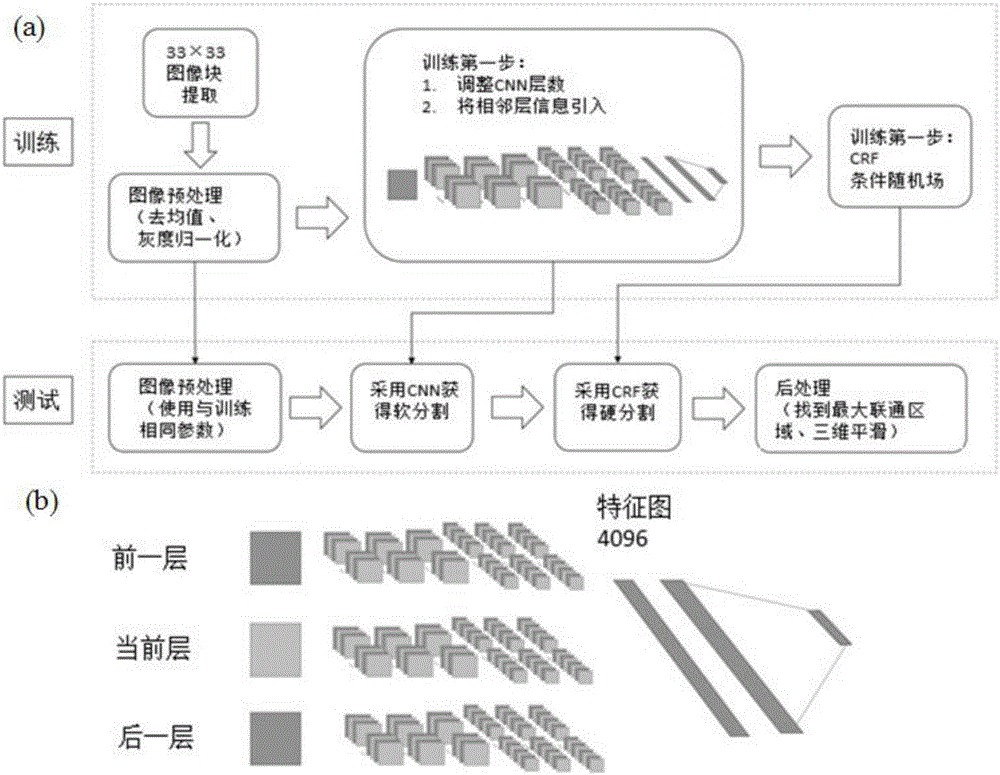
[0061] 1. Firstly, perform operations such as brain removal and grayscale normalization on the original image, manually label 30 cases of 240 pieces of unbiased images as the training set of CNN, and divide the image into 32*32 small blocks and send them to the network for training .
[0062] 2. Use figure 2 The CNN shown segments the image, and subsequently adjusts the segmentation result with a CRF energy random field.
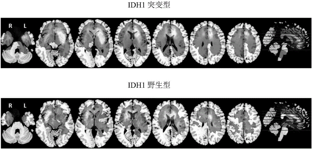
[0063] 3. Map the segmented tumors to the standard brain atlas MN152 with SPM12, superimpose 76 cases of IDH1 mutation and 34 cases of IDH1 wild-type tumors on the standard brain atlas, divide the superposition results into AAL116 partitions, and count the two Distribution of tumoroids on 116 partitions as 116 location features.
[0064] 4. A total of 555 grayscale, shape, texture, and wavelet features shown in Table 1 were extracted, plus 116 positional features, and a...
PUM
 Abstract
Description
Claims
Application Information