
Method and device for optimizing ultrasonic image

An ultrasound and image technology, applied in the medical field, can solve problems such as complicated operations and the inability of ultrasound images to effectively express human body information, and achieve the effect of reducing complexity

Inactive Publication Date: 2017-05-24
SHENZHEN WISONIC MEDICAL TECH CO LTD
View PDF4 Cites 4 Cited by
  • Summary
  • Abstract
  • Description
  • Claims
  • Application Information

AI Technical Summary

Problems solved by technology

[0003] The main purpose of the present invention is to provide a method and device for optimizing ultrasonic images, aiming to solve the technical problems that the existing ultrasonic images cannot effectively express human body information, and users have complicated operations in the process of obtaining ultrasonic images

Method used


Image

  • Method and device for optimizing ultrasonic image
  • Method and device for optimizing ultrasonic image
  • Method and device for optimizing ultrasonic image

Examples


Embodiment Construction

[0056] It should be understood that the specific embodiments described here are only used to explain the present invention, not to limit the present invention.

[0057] The present invention provides a method of optimizing an ultrasound image.

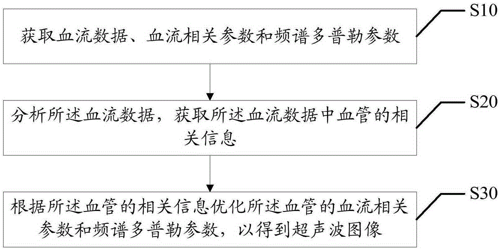
[0058] refer to figure 1 , figure 1 It is a schematic flowchart of the first embodiment of the method for optimizing an ultrasonic image according to the present invention.

[0059] In this embodiment, the method for optimizing an ultrasonic image includes:

[0060] Step S10, acquiring blood flow data, blood flow related parameters and spectral Doppler parameters;

[0061] The Doppler diagnostic instrument generates the excitation pulse signal of each channel through its front-end transistor Transmitor according to the current working parameter conditions and transmits it to its probe Probe, wherein the probe is composed of multiple array elements, and each array element is a connection A transducer with a data channel, the functio...


PUM


Abstract

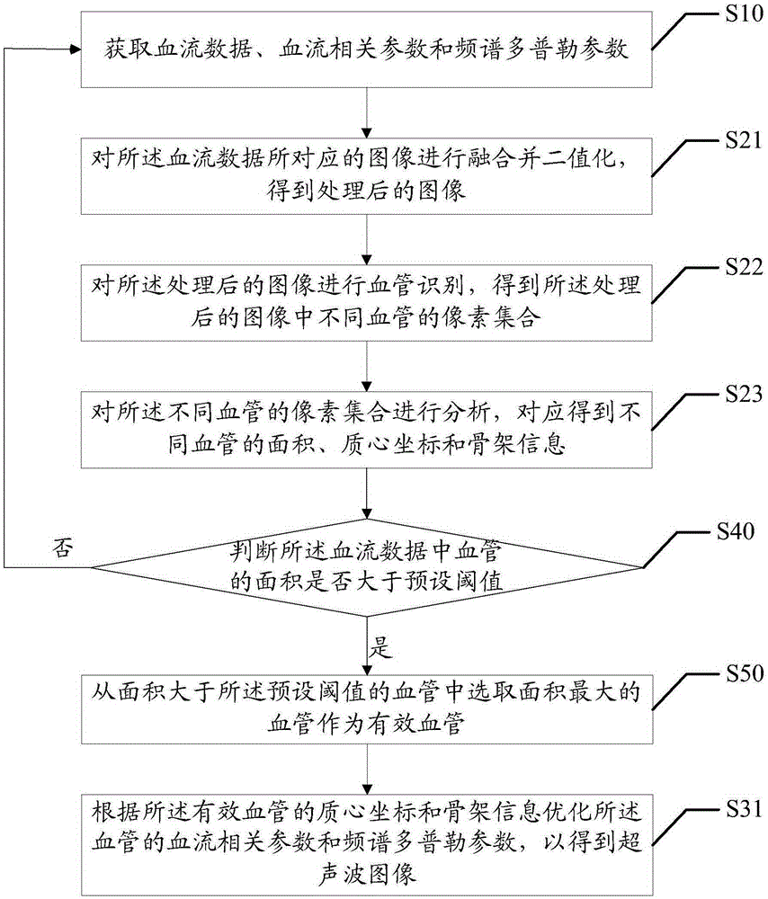
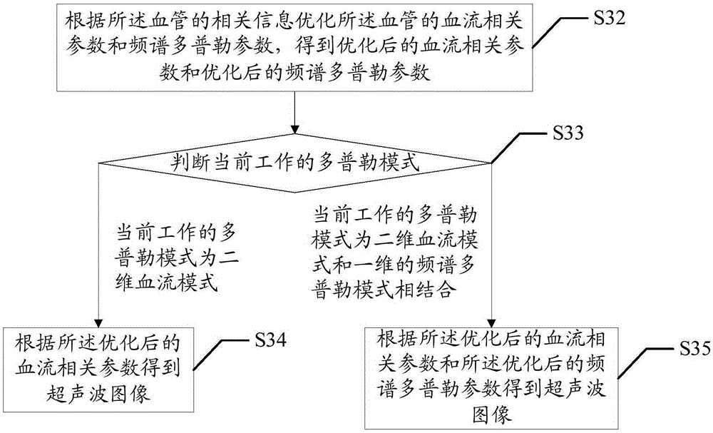
The invention discloses a method for optimizing an ultrasonic image. The method comprises the following steps: acquiring blood flow data, blood flow related parameters and spectral Doppler parameters; analyzing the blood flow data and acquiring related information of blood vessels in the blood flow data; and in accordance with the related information of the blood vessels, optimizing the blood flow related parameters and spectral Doppler parameters of the blood vessels, so as to obtain the ultrasonic image. The invention also discloses a device for optimizing the ultrasonic image. With the application of the method and the device provided by the invention, complexity of user operation is reduced; and meanwhile, the obtained ultrasonic image can effectively express human body information.

Description

technical field [0001] The invention relates to the medical field, in particular to a method and device for optimizing an ultrasonic image. Background technique [0002] The most conventional ultrasonography includes B-mode and Doppler (Doppler) mode, B-mode is used for tissue imaging, and Doppler mode is routinely used for analysis and imaging of blood flow signals in tissues. The two most commonly used modes in Doppler mode are two-dimensional blood flow mode (Color Flow, Color mode) and one-dimensional spectral Doppler mode (Spectral Doppler, also called PW mode), and the latest technology has three-dimensional Doppler mode. Generally, the ultrasound system enters and exits B mode, Color mode and PW mode through its B, C and PW buttons respectively. In routine vascular inspection, users usually use two-dimensional blood flow mode to obtain blood flow information in human tissue. When the user clicks the C button, he can enter and exit the Color mode. When entering the C...

Claims


Application Information

Patent Timeline
IPC IPC(8): A61B8/06G06T7/00
Inventor 姚斌
Owner SHENZHEN WISONIC MEDICAL TECH CO LTD
Learn More