
Nidus and/or organ modeling method and apparatus used for model body making

A modeling method and lesion technology, applied in the field of medical imaging, can solve problems such as difficult clinical services, not to mention the stability of medical phantoms

Active Publication Date: 2017-05-24
上海术理智能科技有限公司
View PDF6 Cites 10 Cited by
  • Summary
  • Abstract
  • Description
  • Claims
  • Application Information

AI Technical Summary

Problems solved by technology

At present, most of the CT phantoms on the market are designed for test equipment rays, and most of them are spherical, cylindrical water phantoms or single PMMA phantoms. There is still a big gap between their shape and bionicity and clinical application; The body is rarely seen in clinical medical centers, and is mostly used in advanced research centers or R&D departments of large equipment companies, and it is also mainly used to standardize mechanical shapes, with material samples of different attenuation densities embedded inside, mainly used for system debugging and testing, it is difficult to truly serve clinical
[0003] At present, there is no clinical medical phantom that can accurately reflect the detailed anatomical structure of the human body, lesion / pathological information, and reflect the performance of imaging medical equipment, let alone the stability of the medical phantom (for example, under long-term exposure to x-rays) and cost affordability

Method used


Image

  • Nidus and/or organ modeling method and apparatus used for model body making
  • Nidus and/or organ modeling method and apparatus used for model body making
  • Nidus and/or organ modeling method and apparatus used for model body making

Examples


Embodiment Construction

[0066] Example embodiments will now be described more fully with reference to the accompanying drawings. Example embodiments may, however, be embodied in many forms and should not be construed as limited to the examples set forth herein; rather, these embodiments are provided so that this disclosure will be thorough and complete and will fully convey the concept of example embodiments to those skilled in the art. The described features, structures, or characteristics may be combined in any suitable manner in one or more embodiments. In the following description, numerous specific details are provided in order to give a thorough understanding of embodiments of the present disclosure. However, those skilled in the art will appreciate that the technical solutions of the present disclosure may be practiced without one or more of the specific details being omitted, or other methods, components, devices, steps, etc. may be adopted. In other instances, well-known technical solution...


PUM


Abstract

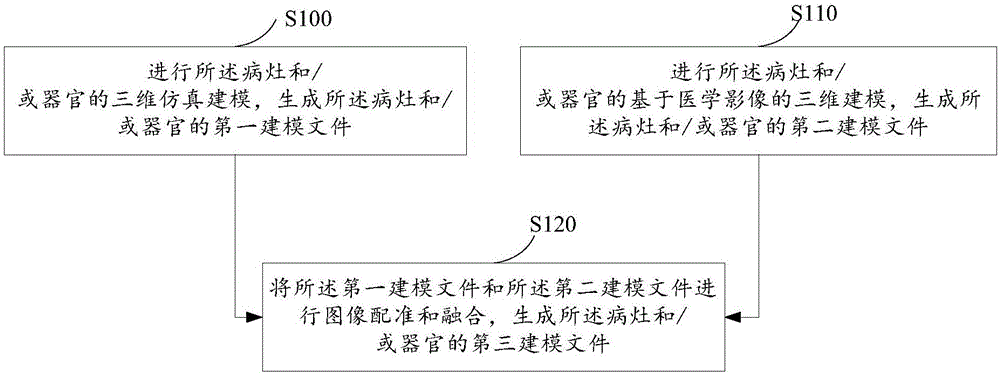
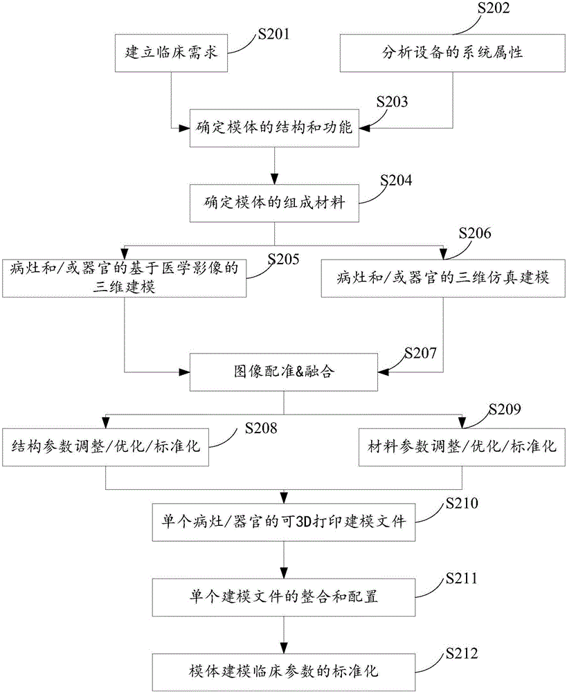
The invention relates to a nidus and / or organ modeling method and apparatus used for model body making. The nidus and / or organ modeling method used for model body making comprises the steps of performing three-dimensional simulation modeling of a nidus and / or an organ to generate a first modeling file of the nidus and / or the organ; performing medical image-based three-dimensional modeling of the nidus and / or the organ to generate a second modeling file of the nidus and / or the organ; and performing image registration and fusion on the first modeling file and the second modeling file to generate a third modeling file of the nidus and / or the organ. According to the method and the apparatus, nidus and / or organ modeling for model body making is realized through the registration and fusion of the three-dimensional simulation modeling and the medical image-based three-dimensional modeling.

Description

technical field [0001] The present disclosure relates to the technical field of medical imaging, in particular, to a lesion and / or organ modeling method, device and electronic equipment for making a phantom. Background technique [0002] At present, the supply of medical phantoms in the market is far from meeting the needs of clinical diagnosis and treatment in terms of technology and clinical popularity. For example, large-scale medical equipment CT and MR are widely used in clinical practice. At present, most of the CT phantoms on the market are designed for test equipment rays, and most of them are spherical, cylindrical water phantoms or single PMMA phantoms. There is still a big gap between their shape and bionicity and clinical application; The body is rarely seen in clinical medical centers, and is mostly used in advanced research centers or R&D departments of large equipment companies, and it is also mainly used to standardize mechanical shapes, with material samples...

Claims


Application Information

Patent Timeline
IPC IPC(8): G06T17/00G06T7/30G06F19/00A61B6/03A61B5/055G09B23/28
CPCA61B5/055A61B6/032A61B6/58A61B2560/0266G06T17/00G06T2207/10081G06T2207/20221G06T2207/30004G06T2207/30096G09B23/28G16H50/50
Inventor 王薇薛华丹
Owner 上海术理智能科技有限公司
Learn More