Microscopy device and method based on smartphone and conical fiber array coupling imaging

A smart phone, tapered optical fiber technology, applied in the field of biomedical detection, can solve the problems of too large optical structure, inconvenient remote data sharing, complex focusing, etc., and achieve the effect of broad prospects, fast enhanced display, and universal practical value

Inactive Publication Date: 2017-05-31
DALIAN UNIV OF TECH
View PDF4 Cites 0 Cited by
  • Summary
  • Abstract
  • Description
  • Claims
  • Application Information

AI Technical Summary

Problems solved by technology

However, these microscopes are still based on the basic principles of optical lens groups, resulting in defects such as excessively large optical structures, complicated focusing, and inability to conveniently realize remote data sharing.

Method used


Image

  • Microscopy device and method based on smartphone and conical fiber array coupling imaging
  • Microscopy device and method based on smartphone and conical fiber array coupling imaging

Examples


Embodiment Construction

[0020] The specific implementation manners of the present invention will be further described below in conjunction with the accompanying drawings and technical solutions.

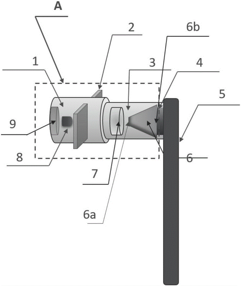
[0021] figure 1 Shows the structural diagram of the microscopic imaging device installed on the smart phone 5 of the present invention, the smart phone 5 may be a mobile device with an Android, ios or windows phone operating system and a built-in camera module. Wherein the sleeve 1 and the sleeve 3 are configured to attach the optical transmission device lens 3, the tapered optical fiber array 6, and the built-in camera 4, and link to the casing of the smart phone.

[0022] figure 1 In the cavity formed by the two-stage detachable focusing sleeve, the imaging device attaches the central point of the sample slide 2, the lens 7, the tapered optical fiber array and the coupling body 6 of the CMOS image sensor of the mobile phone, and Align the center light path. The coupling body has a shorter focal length ...


PUM


Abstract

The invention provides a microscopy device and method based on smartphone and conical fiber array coupling imaging, which belongs to the field of biomedical detection. The high-resolution and portable micro-imaging device and the processing method used for further image enhancement display are designed. According to the invention, the device and method can be widely applied to blood cell pathology detection in biomedicine, further can realize high-definition imaging and sample morphology video tracking, overcome the defects of large volume, cumbersome focusing, complex image transmission step and the like of a traditional optical microscope, and have good application prospects.

Description

technical field [0001] The invention relates to the field of biomedical detection, and more specifically relates to a microscopic device and method based on coupled imaging of a smart phone and a tapered optical fiber array. Background technique [0002] Optical microscopes are widely used in biomedical inspections. However, their large size, high cost, and complex focusing limit their applications in many fields and regions. The expensive lens group especially limits the popularity of microscopes. In order to overcome these problems, hand-held and miniaturized microscopes have developed vigorously in recent years, and have gradually moved from scientific research to practical fields. However, these microscopes are still based on the basic principles of optical lens groups, resulting in defects such as excessively large optical structures, complicated focusing, and inability to conveniently realize remote data sharing. Contents of the invention [0003] In view of the abo...

Claims


Application Information

Patent Timeline
Patent Type & Authority Applications(China)
IPC IPC(8): G02B21/36
CPCG02B21/361G02B21/365
Inventor 喻言王伟明黄辉欧进萍
Owner DALIAN UNIV OF TECH
Why Eureka
Try Eureka