A method for bone marrow fluid cell segmentation based on deep learning

A deep learning and cell technology, applied in the fields of biomedical image processing and computer applications, can solve the problem of inaccurate segmentation results, achieve the effects of accurate segmentation results, improve efficiency, and simplify the calculation process

Active Publication Date: 2019-08-30
杭州华卓信息科技有限公司
View PDF7 Cites 0 Cited by
  • Summary
  • Abstract
  • Description
  • Claims
  • Application Information

AI Technical Summary

Problems solved by technology

However, the segmentation results of such methods are not accurate enough when the cells are poorly stained or the cells are adherent.

Method used


Image

  • A method for bone marrow fluid cell segmentation based on deep learning
  • A method for bone marrow fluid cell segmentation based on deep learning
  • A method for bone marrow fluid cell segmentation based on deep learning

Examples


specific Embodiment

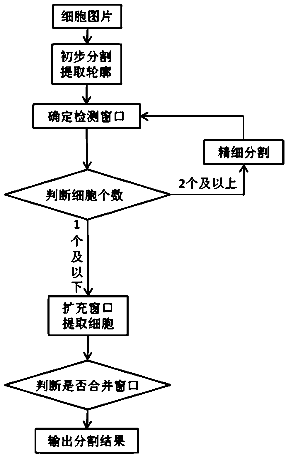
[0092] like figure 1 As shown, the embodiment of the present invention provides a method for segmenting bone marrow fluid cells based on deep learning, which can be implemented by the following steps:

[0093] Step (1), preliminarily estimate the position of the cell nucleus. First, with the vector x i =(r i , g i , b i , r i -g i , b i -g i ) T Represents each pixel in the image, where (r i , g i , b i ) are the RGB components of the pixel, respectively. Use the k-means algorithm to divide these vectors into three categories, and get the center vector μ l =(u 1 , u 2 , u 3 , u 4 , u 5 ,) T ,l=1,2,3. make

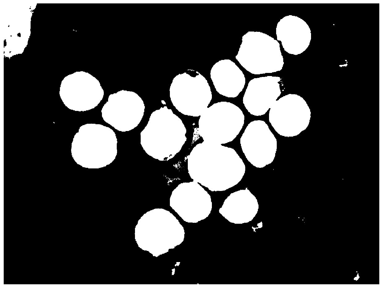
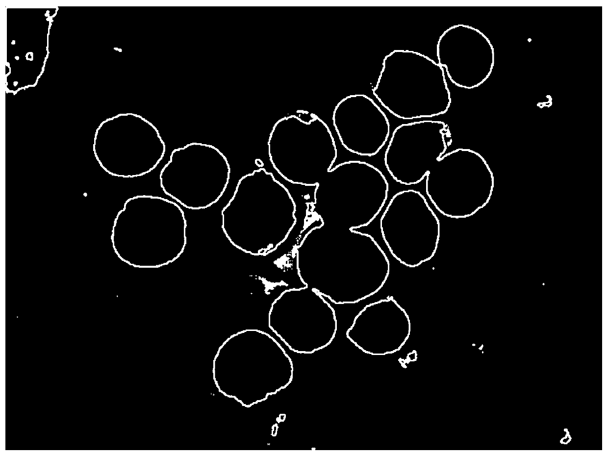
[0094] Compute the class representing the nucleus Create a mask map I (such as figure 2 ), the estimated nucleus position is white, and the rest are black. Extract all contours that mask the white foreground of Figure I (such as image 3 ). Further, calculate the perimeter and area of ​​all contours, that is, the number of pixels on the contour...


PUM


Abstract

The invention discloses a marrow fluid cell partition method based on depth study, and relates to the field of biological medical image treatment and computer application. Compared with the prior art, the marrow fluid cell partition method provided by the embodiment is realized by a clustering algorithm and a depth study method; the method is direct and easy to practice; through an automatic evaluation system of the partitioning quality, the partitioning result is more exact; the component characteristic of the HSV image is fully used, and the unique regional growth method is designed, and the calculation process is greatly simplified; the efficiency is improved.

Description

technical field [0001] The present invention relates to the field of biomedical image processing and computer application, in particular to a method for segmenting bone marrow fluid cells based on deep learning. Background technique [0002] Blood cytology examination of bone marrow fluid is of great significance in the diagnosis of some blood diseases. For the diagnosis of blood diseases, although there are many methods such as fluorescence microscope, phase contrast microscope, electron microscope, molecular biology, cytochemistry, cytogenetics, immunology and biopsy, the examination of cell morphology is still the most basic, The most commonly used diagnostic tool. Most blood diseases can be correctly diagnosed only by cytological examination combined with clinical data. In pathological conditions, particularly in cases of acute leukemia, cell morphology can become malformed. Even experienced hematologists cannot accurately identify these atypical cells. At present, bo...

Claims


Application Information

Patent Timeline
Patent Type & Authority Patents(China)
IPC IPC(8): G06T7/12G06T7/13G06M11/00G06T7/11
Inventor 毛嘉昀居斌李兰娟李谭伟
Owner 杭州华卓信息科技有限公司
Why Eureka
Try Eureka