A Feature Extraction Method of Lightweight Incisional Hernia Mesh 3D Ultrasound Image

An image feature extraction, three-dimensional ultrasound technology, applied in the field of image processing, can solve the problems of weak echo signal, insufficient high echo image, small residual foreign matter, etc., to achieve the effect of improving robustness

CN106875409BInactive Publication Date: 2019-06-21YUNNAN UNIV
1 Cites 0 Cited by

Patent Information

Authority / Receiving Office
CN · China
Patent Type
Patents(China)
Current Assignee / Owner
Publication Date
2019-06-21
Estimated Expiration
Not applicable · inactive patent

Smart Images

  • Figure 1
    Figure 1
  • Figure 2
    Figure 2
  • Figure 3
    Figure 3
Patent Text Reader

Abstract

The invention belongs to the technical field of image processing, in particular to a method for extracting features of a light-weight three-dimensional ultrasonic image of an incisional hernia patch. The present invention first uses the texture feature extraction algorithm to automatically quantify and extract the relevant texture feature parameters of the area to be classified in the three-dimensional volume of interest (VOI) of the automated three-dimensional breast ultrasound (ABUS) image, so as to distinguish the patch from the fascia; and then Aiming at the problem that the two-dimensional texture parameters are sensitive to spatial transformations such as curling and shrinkage of the incisional hernia mesh, three-dimensional texture parameters and three-dimensional position parameters are introduced to improve the robustness of the light-weight patch classification and recognition algorithm; finally, the class The distance algorithm and the sequential forward search method are used for feature selection. The method of the invention has good feature selection effect and high efficiency, can effectively improve the classification accuracy of the light-weight incision hernia patch three-dimensional ultrasonic image, and is convenient for automatic classification and identification.
Need to check novelty before this filing date? Find Prior Art

Description

technical field

[0001] The invention belongs to the technical field of image processing, and in particular relates to a method for extracting features of a lightweight three-dimensional ultrasonic image of an incisional hernia patch. Background technique

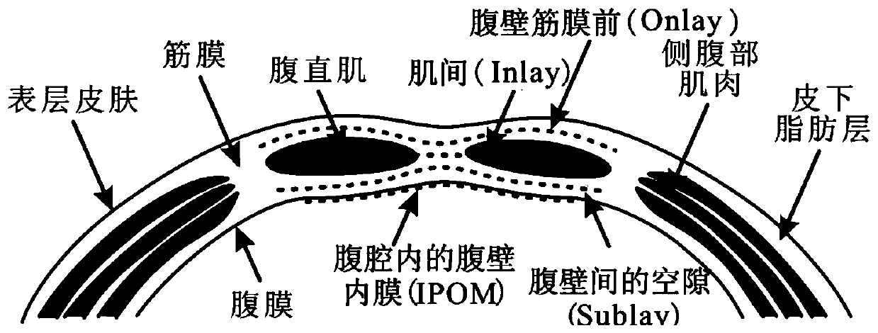
[0002] The thickness of light-weight patches is usually less than 0.5mm, the braided hole diameter is more than 3mm, and the residual foreign matter is very small, resulting in weak echo signals, and the linear hyperechoic images in the imaging results are not obvious enough. At the same time, in abdominal wall herniorrhaphy, lightweight mesh can be placed in the front of the abdominal fascia (Onlay), between the muscles (Inlay), the space between the fascia and the peritoneum (Sublay), and the intraperitoneal wall of the abdominal cavity (IPOM). ) four positions related to the anatomical structure of the abdominal wall (such as figure 2 shown), while all implantation locations were relatively close to the fascial tissue....

Examples

Embodiment Construction

[0091] The present invention will be specifically described below in conjunction with the accompanying drawings and specific implementation methods. Obviously, the described embodiments are only some of the embodiments of the present invention, not all of them. Based on the embodiments of the present invention, all other embodiments obtained by persons of ordinary skill in the art without making creative efforts belong to the protection scope of the present invention.

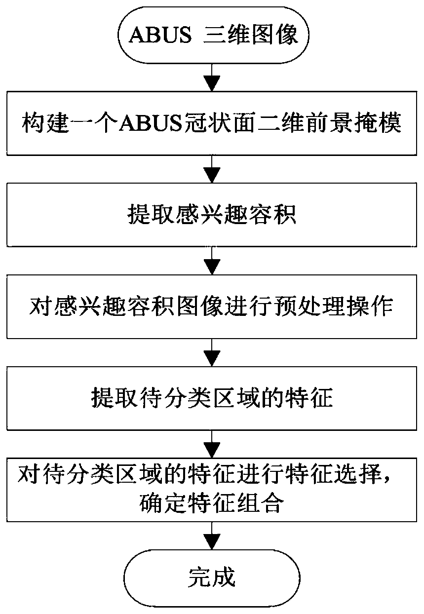
[0092] Refer to attached figure 1 , the light-weight incisional hernia mesh 3D ultrasonic image feature extraction method of the technical solution includes the following steps:

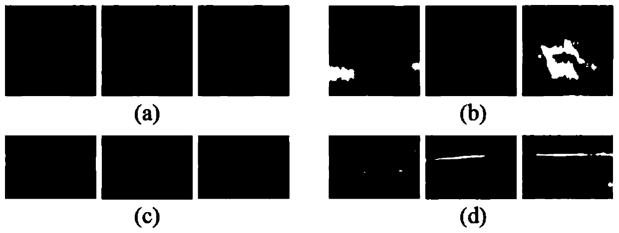
[0093] S1: Construct a 2-D foreground mask of the ABUS coronal plane, the experimental results are as follows Figure 7 as shown in (a);

[0094] (1) All ABUS coronal slices located at 0.5 to 0.9 times the total depth of scan C 1 -C n removed, for all C 1 -C n Pixels at the same position in the image are averaged to obtain a co...