

Lightweight incisional hernia patch three-dimensional ultrasonic image feature extraction method
An image feature extraction and three-dimensional ultrasound technology, applied in the field of image processing, can solve problems such as weak echo signal, small amount of foreign matter residue, and insufficient high echo image, and achieve the effect of improving robustness
- Summary
- Abstract
- Description
- Claims
- Application Information
AI Technical Summary
Problems solved by technology
Method used
Image

Examples
Embodiment Construction
[0091] The present invention will be specifically described below in conjunction with the accompanying drawings and specific implementation methods. Obviously, the described embodiments are only some of the embodiments of the present invention, not all of them. Based on the embodiments of the present invention, all other embodiments obtained by persons of ordinary skill in the art without making creative efforts belong to the protection scope of the present invention.
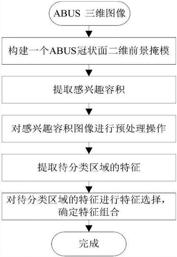
[0092] Refer to attached figure 1 , the light-weight incisional hernia mesh 3D ultrasonic image feature extraction method of the technical solution includes the following steps:
[0093] S1: Construct a 2-D foreground mask of the ABUS coronal plane, the experimental results are as follows Figure 7 as shown in (a);
[0094] (1) All ABUS coronal slices located at 0.5 to 0.9 times the total depth of scan C 1 -C n removed, for all C 1 -C n Pixels at the same position in the image are averaged to obtain a co...
PUM
 Abstract
Description
Claims
Application Information