
Panel PET imaging device with window

An imaging device and a flat-panel technology, applied in the field of medical devices, can solve problems such as insufficient and inability to locate and scan the operation space in real time, and achieve the effects of improving accuracy, improving radiotherapy effects, and avoiding positioning errors.

Active Publication Date: 2017-07-07
RAYCAN TECH CO LTD SU ZHOU
View PDF5 Cites 0 Cited by
  • Summary
  • Abstract
  • Description
  • Claims
  • Application Information

AI Technical Summary

Problems solved by technology

[0011] The purpose of the present invention is to provide a flat-panel PET imaging device with a window, so as to solve the problems in the prior art that real-time positioning and scanning cannot be performed during radiotherapy navigation and the operating space is insufficient

Method used


Image

  • Panel PET imaging device with window
  • Panel PET imaging device with window
  • Panel PET imaging device with window

Examples


Embodiment Construction

[0030] The present invention will be further described below in conjunction with specific embodiments. It should be understood that the following examples are only used to illustrate the present invention and not to limit the scope of the present invention.

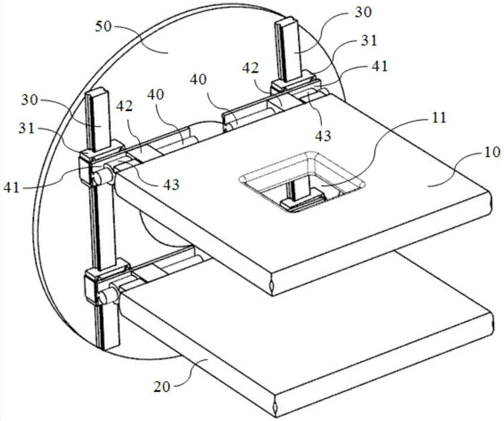
[0031] figure 2 It is a three-dimensional schematic diagram of a flat-panel PET imaging device with a window according to an embodiment of the present invention. figure 2 It can be seen that the flat PET imaging device provided by the present invention includes a first flat plate 10, a second flat plate 20, and a supporting mechanism. The first flat plate 10 and the second flat plate 20 are arranged in parallel and fixed by the supporting device. Specifically, the supporting device includes a first guide rail 30, a second guide rail 40, and a rotating bracket 50, wherein two parallelly arranged first rails 30 are arranged on the same side of the annular plate-shaped rotating bracket 50, and each first rail 30 Two first sli...


PUM


Abstract

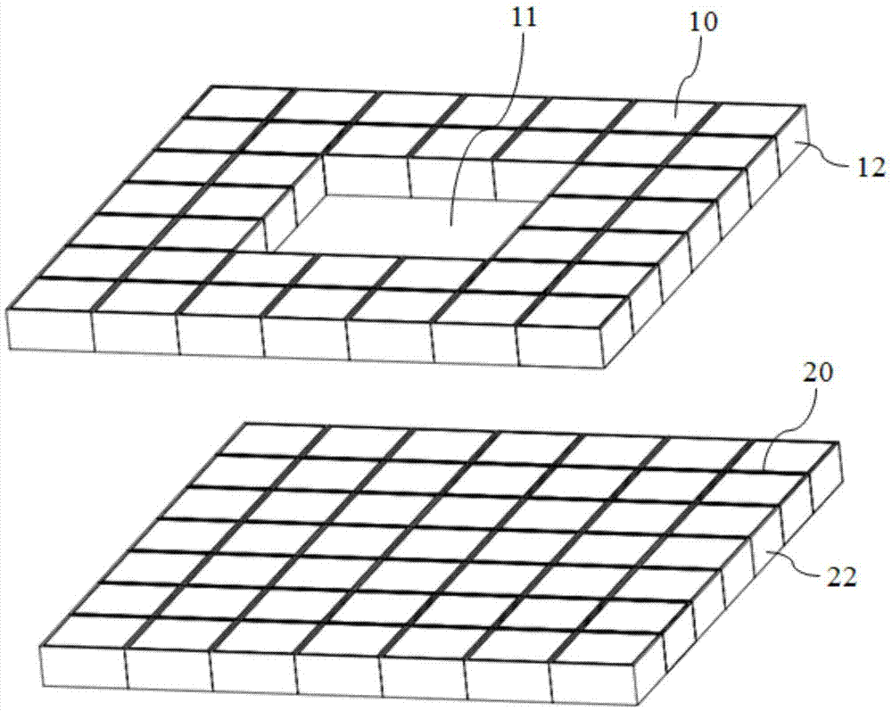
The invention provides a panel PET imaging device with a window. The device comprises a first panel, a second panel and a support device. The first panel is plate-shaped and made by orderly arrangement of multiple PET detectors. The first panel is provided with at least one window. The second panel is plate-shaped and made by orderly arrangement of multiple PET detectors and parallel to the first panel. The first panel and the second panel are fixed on the support device. The panel PET imaging device with the window has the following beneficial effects: by arranging the window on a panel PET, real-time location and scanning effect is ensured while space is provided for radiation oncology and other operation; and real-time diagnoses and positioning navigation can be achieved without affecting treatment operation.

Description

Technical field [0001] The present invention relates to a medical device, and more particularly to a flat-panel PET imaging device with a window. Background technique [0002] Cancer is currently one of the most threatening diseases to human life and health. Surgery, radiotherapy and chemotherapy are commonly used as treatment methods for cancer. For radiotherapy, it uses radioisotope rays, ordinary X-rays generated by X-ray treatment machines, high-energy X-rays generated by accelerators, electron beams generated by various accelerators, protons, fast neutrons, and heavy particles for cancer treatment. As an important cancer treatment method, how to improve the efficacy of radiotherapy is the focus of research. Generally, optimizing the radiation dose distribution can significantly improve the curative effect of radiotherapy, and the key to optimizing the radiation dose distribution is to accurately obtain the physiological and biochemical information of the tumor and its surro...

Claims


Application Information

Patent Timeline
Patent Type & Authority Applications(China)
IPC IPC(8): A61B6/12A61B6/03A61B6/02A61B6/00
CPCA61B6/025A61B6/037A61B6/12A61B6/4435A61B6/44A61N2005/1052A61N5/1049A61B6/4266G01T1/2985
Inventor 李炳轩肖鹏郭彦彬谢庆国
Owner RAYCAN TECH CO LTD SU ZHOU
Learn More