Image segmentation method based on multi-supervision full-convolution neural network

A convolutional neural network and image segmentation technology, applied in the field of medical image processing, can solve the problems of poor segmentation efficiency, large amount of calculation, and insufficient feature extraction, and achieve the effect of reducing segmentation errors, speeding up training speed, and improving segmentation accuracy.

Inactive Publication Date: 2017-09-15
UNIV OF SCI & TECH OF CHINA
View PDF2 Cites 103 Cited by
  • Summary
  • Abstract
  • Description
  • Claims
  • Application Information

AI Technical Summary

Problems solved by technology

[0011] The technical problem to be solved by the present invention is: to overcome the problems of the existing segmentation methods such as insufficient feature extraction, strong feature subjectivity, large amount of calculation, poor robustness, and poor segmentation efficiency, and provide a multi-supervised fully convol...

Method used


Image

  • Image segmentation method based on multi-supervision full-convolution neural network
  • Image segmentation method based on multi-supervision full-convolution neural network
  • Image segmentation method based on multi-supervision full-convolution neural network

Examples


Embodiment Construction

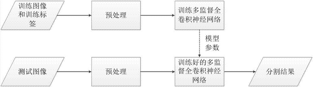
[0025] Such as figure 1 Shown, the segmentation method of a kind of multi-supervision fully convolutional neural network of the present invention, specific technical details are as follows:

[0026] (1) Collect CT images of osteosarcoma and preprocess the images;

[0027] First, anisotropic diffusion filter algorithm is used to denoise the input image, and then the denoised image is standardized to obtain the normalized image.

[0028] (2) Train a multi-supervised fully convolutional neural network model.

[0029] The images normalized in the first step and the labeled images are input into a multi-supervised fully convolutional neural network for training.

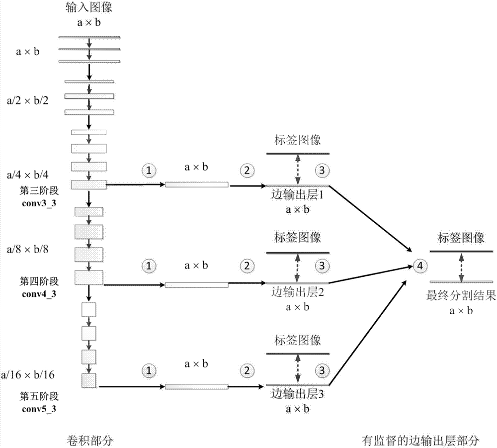
[0030] 1) Multi-supervised fully convolutional network structure.

[0031] Such as figure 2 As shown, the multi-supervised fully convolutional neural network consists of two parts: the convolution part and the supervised edge output layer network part. The original network structure of the vgg-16 model before conv5_3 ...


PUM


Abstract

The invention relates to an image segmentation method based on a multi-supervision full-convolution neural network. According to the invention, further improvement is carried out based on the full convolution neural network, so a novel network structure is provided. The network structure has three edge output layers with supervision which are capable of guiding a network to learn multi-scale features and allowing the network to acquire local features and global features of images. Meanwhile, in order to keep context information in the images, in the upper sampling parts of the network, upper sampling is performed on output feature images by sue of multiple feature channels. Finally, a fusion layer with the weight is used for fusing the classification results of the multiple edge output layers, so the final image segmentation result is obtained. The method is characterized by high segmentation precision and quick segmentation speed. In osteosarcoma CT data segmentation, the DSC coefficient of the acquired segmentation result by use of the provided algorithm reaches 86.88%, which is higher than that of a traditional FCN algorithm.

Description

technical field [0001] The invention relates to an image segmentation method based on a multi-supervised full convolutional neural network, which belongs to the field of medical image processing. Background technique [0002] Computed tomography (Computed Tomography, CT) is a commonly used medical imaging method in the diagnosis and treatment of osteosarcoma. It can accurately segment the tumor lesion area from the CT image of osteosarcoma, and it is necessary for the formulation of preoperative neoadjuvant chemotherapy and radiotherapy plan, as well as the surgical treatment. The evaluation of the curative effect of post-radiotherapy and chemotherapy all plays a crucial role. There is an urgent need for automatic segmentation of tumor regions in clinical practice. [0003] With the development of computer-aided diagnosis technology, researchers have done a lot of outstanding work on the automatic segmentation of osteosarcoma images. In general, these osteosarcoma image se...

Claims


Application Information

Patent Timeline
IPC IPC(8): G06T7/12G06N3/08G06K9/62
CPCG06N3/084G06T7/12G06T2207/10081G06T2207/20081G06T2207/20084G06T2207/30096G06F18/214G06F18/24G06F18/254
Inventor 黄林邱本胜高欣
Owner UNIV OF SCI & TECH OF CHINA
Why Eureka
Try Eureka