Three-dimensional blood vessel segmentation method based on symmetric matching filter group and regional growth

A matched filter and region growing technology, applied in the field of medical image processing, can solve the problems of blood vessel mis-segmentation and over-segmentation, affecting the segmentation effect, etc., and achieve the effects of suppressing response, enhancing display and segmentation, and improving contrast.

Active Publication Date: 2017-11-24
SOUTHEAST UNIV
View PDF4 Cites 10 Cited by
  • Summary
  • Abstract
  • Description
  • Claims
  • Application Information

AI Technical Summary

Problems solved by technology

[0003] Since the main part of the three-dimensional blood vessel in the CTA / MRA image has a high gray value, the main part of the blood vessel can be extracted using the threshold segmentation method. , because the gray value is similar to the background, it is difficult to observe with the naked eye using general imaging tools, and directly using the region growing algorithm with a global fixed threshold to segment blood vessels is prone to mis-segmentation and over-segmentation, which affects the segmentation effect

Method used


Image

  • Three-dimensional blood vessel segmentation method based on symmetric matching filter group and regional growth
  • Three-dimensional blood vessel segmentation method based on symmetric matching filter group and regional growth
  • Three-dimensional blood vessel segmentation method based on symmetric matching filter group and regional growth

Examples


Embodiment Construction

[0034] The present invention will be further described below in conjunction with the accompanying drawings.

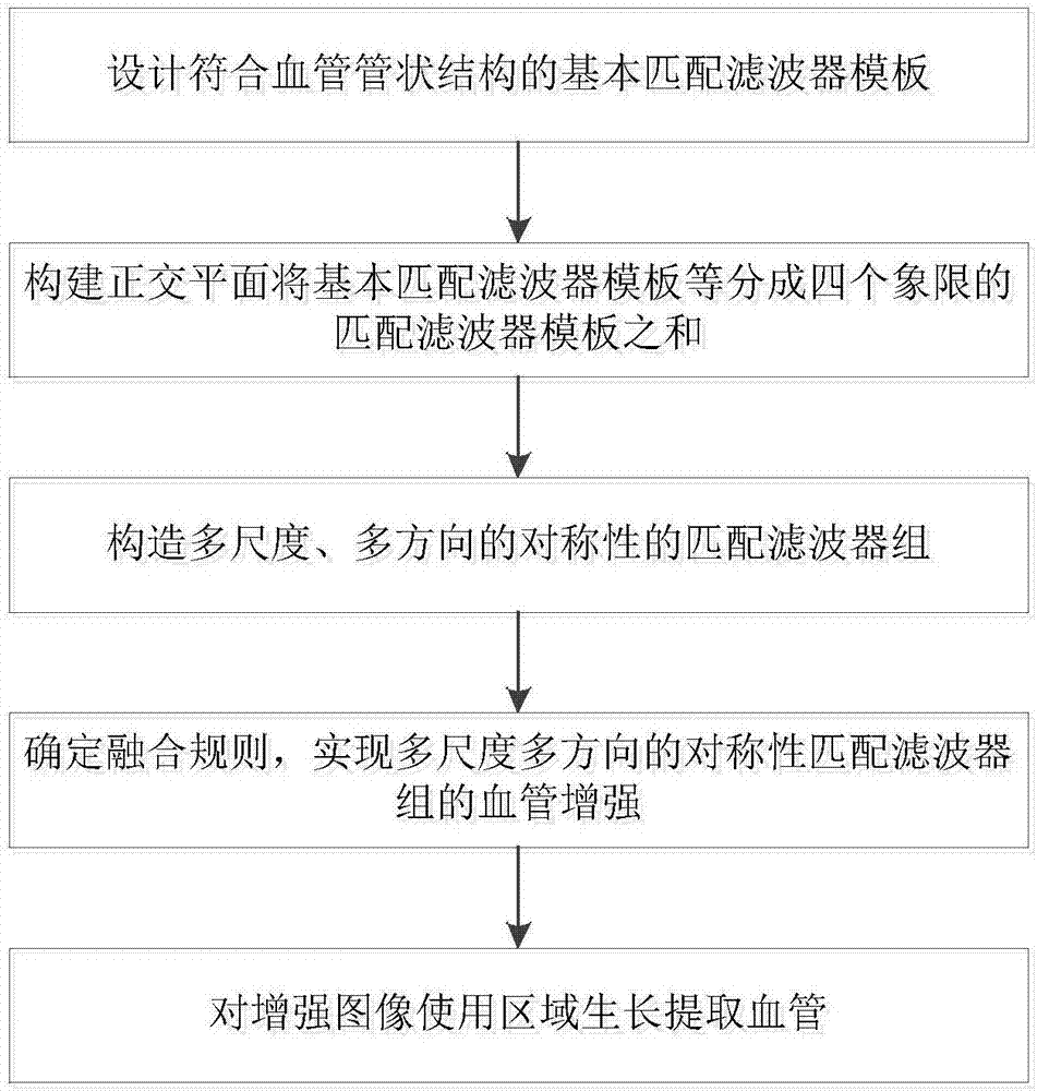
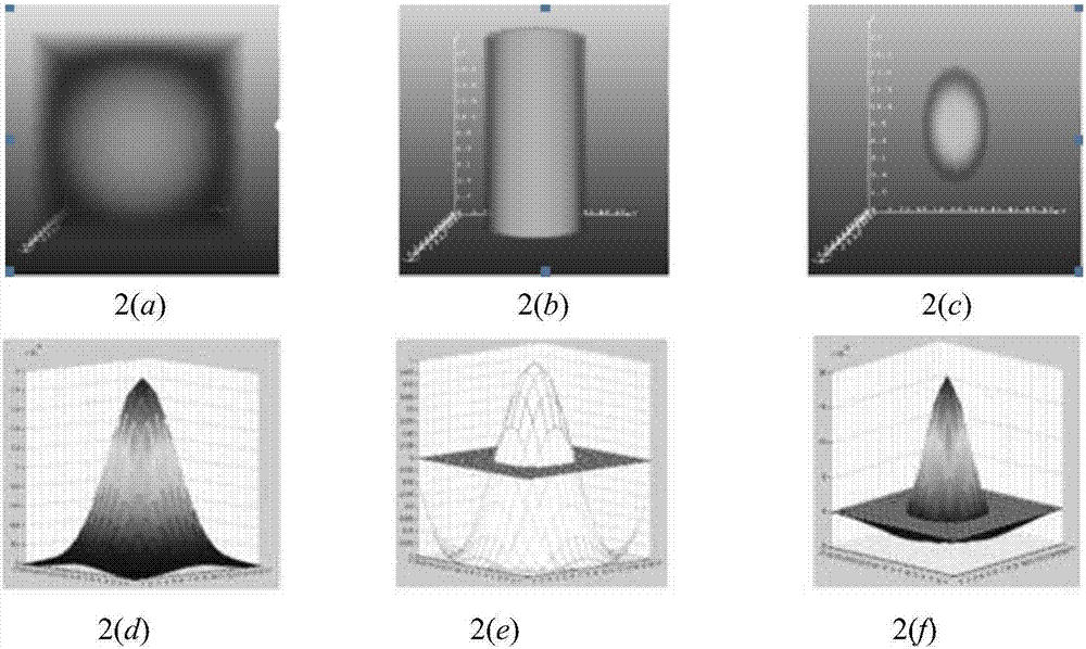
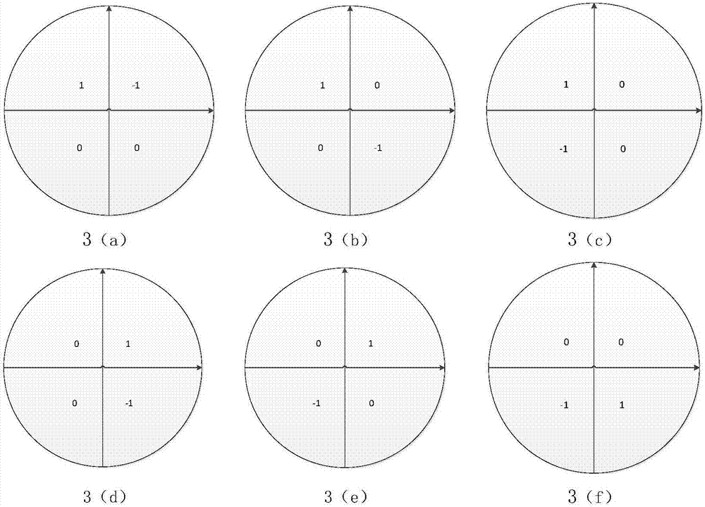
[0035] Such as figure 1 Shown is a 3D blood vessel segmentation method based on symmetrical matched filter banks and region growing. First, starting from the shape characteristics of human 3D blood vessels, a filter function is designed that conforms to the tubular characteristics of blood vessels, and the radial spatial frequency is determined. In the filter template of any scale, form a highlighted tubular structure in the center; then use two orthogonal cut planes in 3D space to divide this tubular structure into four quadrant parts, and assign points to each quadrant in turn The multiplication factor is 1, and the other quadrants are given a dot multiplication factor of 0 to obtain the matched filter partial template corresponding to each quadrant. Then, by adjusting the scale, tubular structures with different diameters are generated, and at the same time, by rot...


PUM


Abstract

The invention discloses a three-dimensional blood vessel segmentation method based on a symmetric matching filter group and regional growth. Firstly, a basic matching filter template of a tubular structure with the satisfaction of a shape characteristic and a gray distribution characteristic of a three-dimensional blood vessel is designed, and two orthogonal planes are used to equally divide a basic matching filter into four quadrants. Then a matching filter group whose direction and scale can be changed is designed, the spatial convolution with a medical image to be enhanced is carried out, the obtainment of a maximum convolution response value is determined as a fusion rule, the blood vessel enhancement of a multi-scale and multi-direction matching filter group is realized, an enhanced image is obtained, and finally the regional growth is used to extract a three-dimensional blood vessel for the enhanced image. According to the method disclosed by the invention, a blood vessel visual effect can be effectively enhanced, the method has a good enhancement effect for a small branch peripheral blood vessel and a blood supply artery close to a lesion tumor especially, at the same time the to suppression of the limbic cortex and other impurities is obvious, thus the contrast of a vessel and a background significantly enhanced, and a solid base is laid for the extraction of a final vessel.

Description

technical field [0001] The invention relates to a three-dimensional blood vessel enhancement and segmentation technology of a CTA image in the field of medical image processing, in particular to a three-dimensional blood vessel segmentation method of a medical image based on a symmetrical matched filter bank and region growing. Background technique [0002] Vascular disease has become one of the most important public health problems. Faced with specific size and complex angiographic images, segmenting blood vessels from them is a key step towards accurate visualization, diagnosis and quantitative analysis of vascular lesions. Blood vessels in different parts of the human body can provide a large amount of information about the relevant tissues of the human body. Their shape (mainly including the diameter of the blood vessel, the bifurcation angle of the blood vessel, and the curvature of the blood vessel) and distribution are important indicators for the diagnosis of related ...

Claims


Application Information

Patent Timeline
Patent Type & Authority Applications(China)
IPC IPC(8): G06T7/11G06T7/136G06T15/00
CPCG06T7/11G06T7/136G06T15/00G06T2207/10012G06T2207/20024G06T2207/30101
Inventor 杨冠羽丁皓宁秀芳舒华忠
Owner SOUTHEAST UNIV
Why Eureka
Try Eureka