Three-dimensional structure for cardiac muscular tissue regeneration and manufacturing method therefor

A three-dimensional structure and tissue technology, applied in tissue regeneration, biochemical equipment and methods, tissue culture, etc., can solve problems such as difficulty in fixing organs, high mortality rate, expensive treatment costs, etc.

Inactive Publication Date: 2017-11-28
T&R BIOFAB
View PDF3 Cites 2 Cited by
  • Summary
  • Abstract
  • Description
  • Claims
  • Application Information

AI Technical Summary

Problems solved by technology

Furthermore, in patients with severe end-stage heart failure, there is no treatment other than heart transplantation or a mechanical left ventricular assist device
But there are problems such as lack of donated organs, difficulty in fixing organs, high mortality rate and expensive treatment costs

Method used


Image

  • Three-dimensional structure for cardiac muscular tissue regeneration and manufacturing method therefor
  • Three-dimensional structure for cardiac muscular tissue regeneration and manufacturing method therefor
  • Three-dimensional structure for cardiac muscular tissue regeneration and manufacturing method therefor

Examples


preparation example 1

[0094] Preparation Example 1: Preparation of the first bioprinting composition

[0095] 1-1: Preparation of acellular extracellular matrix

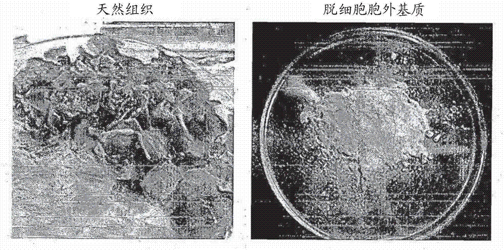
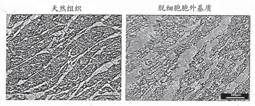
[0096] Acellular extracellular matrix (hereinafter referred to as 'hdECM') was prepared using porcine heart tissue according to the method disclosed in Falguni Pati et al., Nat Commun. 5, 3935 (2014). The prepared hdECM was finally lyophilized and stored frozen until use. Optical micrographs and tissue staining photos are shown in figure 2 middle.

[0097]1-2: Preparation of tissue engineering construct-forming solution in pregel form

[0098] Pour into liquid nitrogen to obtain lyophilized hdECM, and subsequently crush with a mortar and pestle. The obtained hdECM powder (330 mg) and pepsin (33 mg) (P7125, Sigma-Aldrich) were added to a 0.5 M acetic acid aqueous solution (10 ml), followed by stirring at room temperature for 48 hours. The resulting solution was kept below 10°C, and the pH was controlled to about pH7 by adding ribofla...

preparation example 2

[0101] Preparation Example 2: Preparation of the Second Bioprinting Composition

[0102] 2-1: Preparation of acellular extracellular matrix

[0103] Acellular extracellular matrix (hereinafter referred to as 'hdECM') was prepared using porcine heart tissue according to the method disclosed in Falguni Pati et al., Nat Commun. 5, 3935 (2014). The prepared hdECM was finally lyophilized and stored frozen until use. Optical micrographs and tissue staining photos are shown in figure 2 middle.

[0104] 2-2: Preparation of tissue engineering construct-forming solution in pregel form

[0105] Liquid nitrogen was poured into the obtained lyophilized hdECM, and then pulverized with a mortar and pestle. The obtained hdECM powder (330 mg) and pepsin (33 mg) (P7125, Sigma-Aldrich) were added to a 0.5 M acetic acid aqueous solution (10 ml), followed by stirring at room temperature for 48 hours. The resulting solution was kept below 10°C, and the pH was controlled to about pH7 by adding...

Embodiment 1

[0111] Example 1: Preparation of three-dimensional constructs for tissue engineering

[0112] The first bioprinting composition and the second bioprinting composition prepared in Preparation Examples 1 and 2 were used to fabricate a three-dimensional structure.

[0113] Specifically, the polycaprolactone (PCL) backbone was loaded on the syringe (the first syringe) of the multi-head tissue and organ printing system (Jin-Hyung Shim et al., J. Micromech. Microeng. 22085014 (2012)), and heated to About 80°C to melt the polymer. The first bioprinting composition in pregel form obtained in Preparation Example 1 and the second bioprinting composition prepared in Preparation Example 2 were respectively loaded on additional syringes (second and third syringes), and the temperature was maintained below about 10°C. A thin PCL backbone with a linewidth below about 100 μm, a gap of about 300 μm, and a thickness of 120 μm was prepared by applying a pneumatic pressure of about 600 kPa to t...


PUM

PropertyMeasurementUnit
thicknessaaaaaaaaaa
modulusaaaaaaaaaa
thicknessaaaaaaaaaa

Abstract

Provided are a method for manufacturing a three-dimensional structure for cardiac muscular tissue regeneration, and a three-dimensional structure for cardiac muscular tissue regeneration, the method comprising the steps of: forming a three-dimensional structure by printing and crosslinking with a first bioprinting composition and a second bioprinting composition so as to alternately arrange a first bioprinted layer and a second bioprinted layer, wherein the first bioprinting composition contains cardiac progenitor cells and a tissue engineering structure formation solution containing decellularized extracellular matrix and a crosslinking agent, and the second bioprinting composition contains a tissue engineering structure formation solution, mesenchymal stem cells, and a vascular endothelial growth factor; and obtaining a crosslinked-gelled three-dimensional structure by performing thermal gelation on the crosslinked three-dimensional structure. The manufacturing method according to the present invention can uniformly position cardiac progenitor cells in the structure and maintain the viability of cells for a long time by implementing a blood vessel network composed of vascular cells in the structure, thereby remarkably improving the efficiency of cellular delivery into the myocardium.

Description

technical field [0001] The invention relates to a three-dimensional structure for regenerating myocardial tissue and a preparation method thereof. More specifically, the present invention relates to a method for preparing a three-dimensional construct for regenerating myocardial tissue and a three-dimensional construct for regenerating myocardial tissue, the method comprising: a step of forming a three-dimensional construct by printing and cross-linking a first A bioprinting composition and a second bioprinting composition to alternately arrange the first bioprinting layer and the second bioprinting layer, wherein the first bioprinting composition comprises a formed tissue comprising a decellularized extracellular matrix and a cross-linking agent an engineered solution and cardiac progenitor cells, the second bioprinting composition comprising the solution for forming a tissue engineered structure, mesenchymal stem cells, and vascular endothelial growth factor; and gelling the...

Claims


Application Information

Patent Timeline
Patent Type & Authority Applications(China)
IPC IPC(8): C12N5/077A61L27/36C07K14/475
CPCA61L27/3633C07K14/475C12N5/0657B33Y70/00A61L27/36A61F2240/001B33Y10/00B33Y80/00A61L27/367A61L27/3826A61L27/3834A61L27/3891A61L27/3895A61L2430/20C12N5/0697C12N2501/165C12N2502/1329C12N2502/1352C12N2513/00C12N2533/90C12N2537/10
Inventor 曺东佑张珍娥朴薰濬
Owner T&R BIOFAB
Why Eureka
Try Eureka