Method, reagents, and kits for detecting minimal residual disease

A reagent and antibody technology, applied in the field of cancer diagnosis, can solve problems such as hindering the detection of low-frequency malignant cells

Active Publication Date: 2017-12-01
ERASMUS UNIV MEDICAL CENT ROTTERDAM ERASMUS MC
View PDF4 Cites 9 Cited by
  • Summary
  • Abstract
  • Description
  • Claims
  • Application Information

AI Technical Summary

Problems solved by technology

However, it should be noted that a high frequency of normal regenerative cells can interfere with the detection of low frequencies of malignant cells in blood, bone marrow, and other bodily fluids (e.g., cerebrospinal fluid) during and after treatment and after hematopoietic stem cell transplantation

Method used


Image

  • Method, reagents, and kits for detecting minimal residual disease
  • Method, reagents, and kits for detecting minimal residual disease
  • Method, reagents, and kits for detecting minimal residual disease

Examples


Embodiment 1

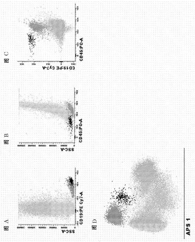
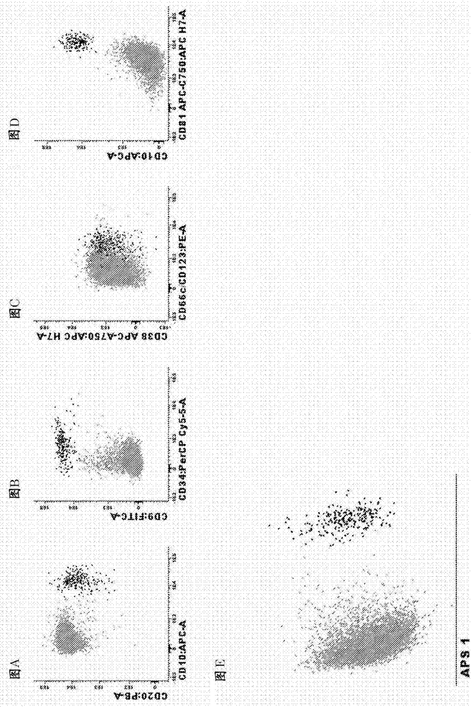
[0096] Example 1. Antibody panels and diagnostic methods for MRD detection in BCP-ALL patients

[0097] Markers for the identification of total B cells and B cell precursors in the bone marrow

[0098] List of relevant identification markers : CD19, CD45

[0099] how to use them : Pre-gating using the CD19 marker is necessary to identify pure B cell populations. To focus on normal B-cell precursors (BCPs), CD45-negative or weakly positive CD45-positive mature B cells can be distinguished from BCPs. In the case of CD19-directed therapy, CD22 can be used instead of CD19. These markers can also be used in combination with side light scatter (SSC) or forward light scatter (FSC) or both FSC and SSC to identify peripheral blood or bone marrow or other types of samples (e.g. bone marrow, tissue biopsy, spinal fluid) B cells in. Of note, additional markers (such as CD10, CD20, CD38, and CD34) (see below) that differentiate normal BCP cells from BCP-ALL cells (see below) can ...

Embodiment 2

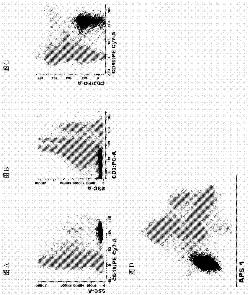
[0114] Example 2. Antibody panels and diagnostic methods for MRD detection in CLL patients

[0115] Markers used to identify total B cells in peripheral blood and bone marrow:

[0116] list of identification markers : CD19, CD3 (markers excluded)

[0117] how to use them : Pre-gating with this marker combination is necessary to identify pure B cell populations and remove T cell / B cell doublets. These markers can also be used in combination with side light scatter (SSC) or forward light scatter (FSC) or both FSC and SSC to identify of B cells. For finer gating to better enrich for CLL cells, both CD5 and CD27 can be used. Markers used to differentiate normal B cells from CLL cells:

[0118] List of markers and most common phenotypic aberrations :

[0119] CD27 : Positive on CLL cells and a small fraction of normal B cells

[0120] CD5 : Positive on CLL cells and a small fraction of normal B cells

[0121] CD79b : Underexpressed on CLL cells compared to norm...

Embodiment 3

[0129] Example 3. Antibody panels and diagnostic methods for MRD detection in MM / PCD patients

[0130] Markers used to identify total plasma cells in bone marrow:

[0131] list of identification markers : CD38, CD138 and CD229

[0132] How to use them:Any combination of the three markers works at any fluorochrome position; any combination of two of the three markers may also be used or in some cases (not all) even one of the three markers alone may be used. The combination is preferably in the following order: 1) CD138 / CD38 / CD229; 2) CD138 / CD38, 3) CD138 / CD229; 4) CD38 / CD229; 5) CD138; 6) CD38); 7) CD229. It should be noted that any of these markers alone and in combination can also be used in combination with side light scatter (SSC) or forward light scatter (FSC) or both FSC and SSC to identify bone marrow or other types of samples (e.g. peripheral blood, Plasma cells in tissue biopsy, spinal fluid).

[0133] Markers to differentiate normal from clonal / malignant pla...


PUM


Abstract

The invention relates to the field of minimal residual disease (MRD) diagnostics, which is progressively more applied for the evaluation of treatment effectiveness in patients with a hematological malignancy, such as B-cell precursor acute lymphoblastic leukemia (BCP-ALL), B-cell chronic lymphocytic leukemia (B-CLL), and multiple myeloma (MM). Provided are unique reagent compositions with carefully selected and thoroughly tested combinations of antibodies, for >=8 color flow cytometric stainings as well as for 10-color and 12-color flow cytometric stainings, which can reach sensitivities of at least 10<-4>, even down to 10<-5>. Also provided are diagnostic kits and methods for detecting MRD.

Description

[0001] This application is a divisional application of the Chinese patent application with application number 201380037624.4. The original application is the PCT international application PCT / NL2013 / 050420 filed on June 14, 2013, which entered the Chinese national phase on January 14, 2015. technical field [0002] The present invention relates to the field of cancer diagnostics, more particularly to means and methods for monitoring disease progression during and after treatment or for detecting minimally disseminated disease. Cytostatic or cytotoxic therapy induces remission in most patients with lymphoid malignancies. However, many of these patients relapse. Clearly, existing cytosuppressive or cytotoxic regimens do not kill all malignant cells in these relapsed patients, although they achieved a so-called complete remission according to cytomorphological criteria. Since the limit of detection of the cytomorphological technique is no lower than 1% to 5% of malignant cells, ...

Claims


Application Information

Patent Timeline
Patent Type & Authority Applications(China)
IPC IPC(8): G01N33/574G01N15/14
CPCG01N33/57426G01N15/14G01N33/57484G01N2015/1006A61K38/164A61K38/1716A61K39/025A61K39/09
Inventor 雅各布斯·约翰内斯·玛丽亚·万东恩何塞·阿尔贝托·奥尔方德马托斯克瑞亚埃韦尔朱安·亚历杭德罗·弗洛雷斯蒙特罗茱莉亚·玛丽亚·阿尔梅达帕拉文森特·亨利库斯·约翰内斯·万德费尔登塞巴斯蒂安·博埃特赫尔朗托尼·威廉·朗格拉克埃斯特尔·迈斯特日科娃托马斯·什切潘斯基马蒂亚斯·里特根保罗·约根·蒙泰罗达席尔瓦卢西奥
Owner ERASMUS UNIV MEDICAL CENT ROTTERDAM ERASMUS MC
Why Eureka
Try Eureka