Anti-idiotypic antibody detection method

An anti-idiotypic antibody and detection method technology, applied in the biological field, can solve the problems of easy false positives and low detection sensitivity of anti-idiotypic antibodies, and achieve the advantages of eliminating the need for enzyme-labeled secondary antibodies, sensitive reactions, and eliminating false positives Effect

Active Publication Date: 2019-08-23
SHENZHEN CENT FOR DISEASE CONTROL & PREVENTION
View PDF2 Cites 0 Cited by
  • Summary
  • Abstract
  • Description
  • Claims
  • Application Information

AI Technical Summary

Problems solved by technology

[0005] The purpose of the present invention is to overcome the above-mentioned deficiencies of the prior art and provide a method for detecting anti-idiotypic antibodies, which aims to solve the technical problems of low detection sensitivity of existing anti-idiotypic antibodies and prone to false positives

Method used


Image

  • Anti-idiotypic antibody detection method
  • Anti-idiotypic antibody detection method
  • Anti-idiotypic antibody detection method

Examples


Embodiment 1

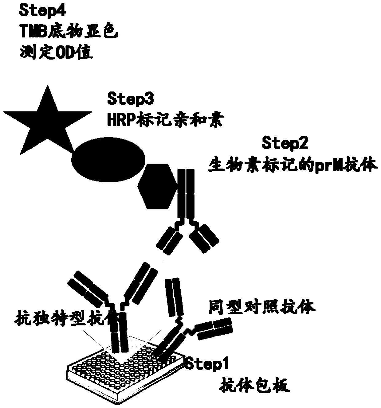
[0053] 1. Coated titer plate

[0054] Add 1 mg / mL anti-idiotypic antibody sample to coating buffer at a ratio of 1:1000, dilute it to EP tube; immediately add 100 μL / well to the microtiter plate, and incubate at 4°C overnight or at room temperature for 2 hours. Negative control: the isotype control antibody is also diluted with coating buffer, and added to the microtiter plate at 0.1ug / well.

[0055] 2. closed

[0056] Pour off the coating buffer in the ELISA plate, add 150 μL of blocking solution to each well, block at room temperature for 1 hour, and wash 4 times with PBST.

[0057] 3. Detection of antibodies

[0058] Dilute the biotin-labeled prM monoclonal antibody (biotin-prM) with sample diluent so that the concentration is 1 μg / ml, add 0.1 ml to each well, and incubate the plate at room temperature for 60 min, or at 37 °C for 30 min. Then wash 4 times with PBST.

[0059] 4. Streptavidin-HRP

[0060] Streptavidin-HRP (Streptavidin-HRP) was diluted by adding sample d...

Embodiment 2

[0066] 1. Coated titer plate

[0067] Add 1 mg / mL anti-idiotypic antibody sample with coating buffer at a ratio of 1:10000, dilute it into EP tube; immediately add 100 μL / well to the microtiter plate, and incubate at 4°C overnight or at room temperature for 2 hours. Negative control: the isotype control antibody is also diluted with coating buffer, and added to the microtiter plate at 0.1ug / well.

[0068] 2. closed

[0069] Pour off the coating buffer in the ELISA plate, add 150 μL of blocking solution to each well, block at room temperature for 1 hour, and wash 4 times with PBST.

[0070] 3. Detection of antibodies

[0071] Dilute the biotin-labeled prM monoclonal antibody (biotin-prM) with sample diluent so that the concentration is 1 μg / ml, add 0.1 ml to each well, and incubate the plate at room temperature for 60 min, or at 37 °C for 30 min. Then wash 4 times with PBST.

[0072] 4. Streptavidin-HRP

[0073] Streptavidin-HRP (Streptavidin-HRP) was diluted by adding sam...


PUM

PropertyMeasurementUnit
concentrationaaaaaaaaaa

Abstract

The invention belongs to the biotechnical field, and concretely relates to an anti-idiotype antibody detection method. The method comprises the following steps: respectively diluting a sample solution and a negative contrast solution with a coating buffer solution to obtain a diluted sample solution and a diluted negative contrast solution; dividing tiny wells in an enzyme label plate into sample wells and negative wells, adding the diluted sample solution to the sample wells, adding the diluted negative contrast solution to the negative wells, carrying out standing treatment, and pouring the diluted sample solution and the diluted negative contrast solution; sequentially adding a blocking solution to the sample wells and the negative wells, carrying out a blocking reaction, adding a sample dilutent diluted biotin-monoclonal antibody and streptavidin-HRP to carry out a connection reaction, carrying out a TMB substrate color developing agent to carry out a color development reaction, adding a stopping solution, and detecting the optical density in the sample wells and the negative wells by an enzyme-linked immunometric meter to determine whether an anti-idiotype antibody corresponding to the monoclonal antibody exists in the sample solution or not. The method has the advantages of simple steps, sensitivity enhancement, and no false positive error.

Description

technical field [0001] The invention belongs to the field of biotechnology, and in particular relates to an anti-idiotypic antibody detection method. Background technique [0002] Dengue virus is widely prevalent in tropical and subtropical regions of the world. In my country, it is mainly prevalent in Hainan and Guangdong. Four different serotypes of dengue virus can co-circulate in the same area. The prM antibody produced in patients with dengue virus infection for the first time can enhance the infection of heterotypic virus, causing life-threatening symptoms of dengue hemorrhagic fever (DHF) and dengue shock (DSS). [0003] Anti-idiotype antibody (anti-idiotype antibody; AId) is an anti-antibody against a specific epitope group (called idiotype) on the V region of an antibody molecule. AId and the determinant molecule of the original antigen are mutually "internal image" relationship, which can simulate the effect of antigen structure and function, and can be used as a ...

Claims


Application Information

Patent Timeline
Patent Type & Authority Patents(China)
IPC IPC(8): G01N33/569G01N33/535G01N33/577
Inventor 王淼张仁利黄达娜阳帆
Owner SHENZHEN CENT FOR DISEASE CONTROL & PREVENTION
Why Eureka
Try Eureka