Evaluation Method of Collagen Tissue on Gastric Serosa Surface in Resected Specimens of Gastric Cancer
An evaluation method and collagen tissue technology, applied in the field of pathological identification, can solve the problems of insufficient collagen information, limited accuracy, complicated and time-consuming operations, etc., and achieve good scientific research and promotion value, high resolution, and mature technology. Effect
- Summary
- Abstract
- Description
- Claims
- Application Information
AI Technical Summary
Problems solved by technology
Method used
Image

Examples
Embodiment 1
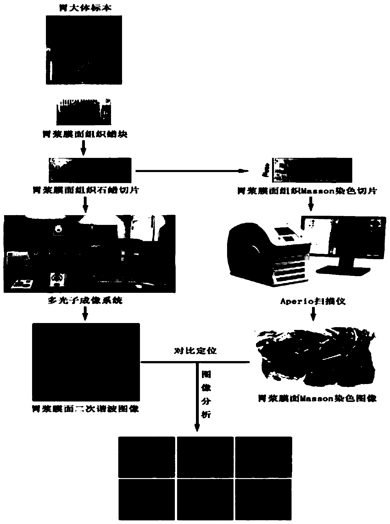
[0055] Such as figure 1 Shown, a method for evaluating collagen tissue on the gastric serosa surface of a gastric cancer resection specimen, the method may further comprise the steps:
[0056] (a) Sample preparation: the gastric serosa surface tissue wax block was prepared after the gastric serosa tissue was separated from the gastric cancer resection specimen, and then a tissue section with a thickness of 3 μm was cut horizontally along the gastric serosa surface of the wax block, and the gastric subserosa Embedded facing downwards to prepare multiphoton imaging slices to be tested;
[0057] (b) Multiphoton collagen imaging: perform second harmonic imaging on the multiphoton imaging slice to be measured in step (a) to obtain a multiphoton collagen imaging image; the output power of the single-channel detector is 1.5W, and the imaging wavelength is 800nm , the receiving wavelength is 390nm, and the imaging multiple is 10 times;
[0058] (c) Section staining: perform Masson s...
Embodiment 2
[0061] A method for evaluating collagen tissue on the gastric serosa surface of a gastric cancer resection specimen, the method comprising the following steps:
[0062] (a) Sample preparation: the gastric serosa surface tissue wax block was prepared after the gastric serosa tissue was separated from the resection specimen of gastric cancer, and then a tissue section with a thickness of 8 μm was cut horizontally along the gastric serosa surface of the wax block, and the gastric subserosa Embedded facing downwards to prepare multiphoton imaging slices to be tested;
[0063] (b) Multiphoton collagen imaging: perform second harmonic imaging on the multiphoton imaging slice to be tested in step (a) to obtain a multiphoton collagen imaging image; the output power of the single-channel detector is 1.8W, and the imaging wavelength is 820nm , the receiving wavelength is 410nm, and the imaging multiple is 10 times;
[0064] (c) Section staining: perform Masson staining on the section a...
Embodiment 3
[0067] A method for evaluating collagen tissue on the gastric serosa surface of a gastric cancer resection specimen, the method comprising the following steps:
[0068] (a) Sample preparation: the gastric serosa surface tissue wax block was prepared after the gastric serosa tissue was separated from the resection specimen of gastric cancer, and then a tissue section with a thickness of 5 μm was cut horizontally along the gastric serosa surface of the wax block, and the gastric subserosa Embedded facing downwards to prepare multiphoton imaging slices to be tested;
[0069] (b) Multiphoton collagen imaging: perform second harmonic imaging on the multiphoton imaging slice to be tested in step (a) to obtain a multiphoton collagen imaging image; the output power of the single-channel detector is 1.6W, and the imaging wavelength is 810nm , the receiving wavelength is 400nm, and the imaging multiple is 10 times;
[0070] (c) Section staining: perform Masson staining on the section a...
PUM
| Property | Measurement | Unit |
|---|---|---|
| thickness | aaaaa | aaaaa |
Abstract
Description
Claims
Application Information
 - R&D Engineer
- R&D Manager
- IP Professional
- Industry Leading Data Capabilities
- Powerful AI technology
- Patent DNA Extraction
Browse by: Latest US Patents, China's latest patents, Technical Efficacy Thesaurus, Application Domain, Technology Topic.
© 2024 PatSnap. All rights reserved.Legal|Privacy policy|Modern Slavery Act Transparency Statement|Sitemap