

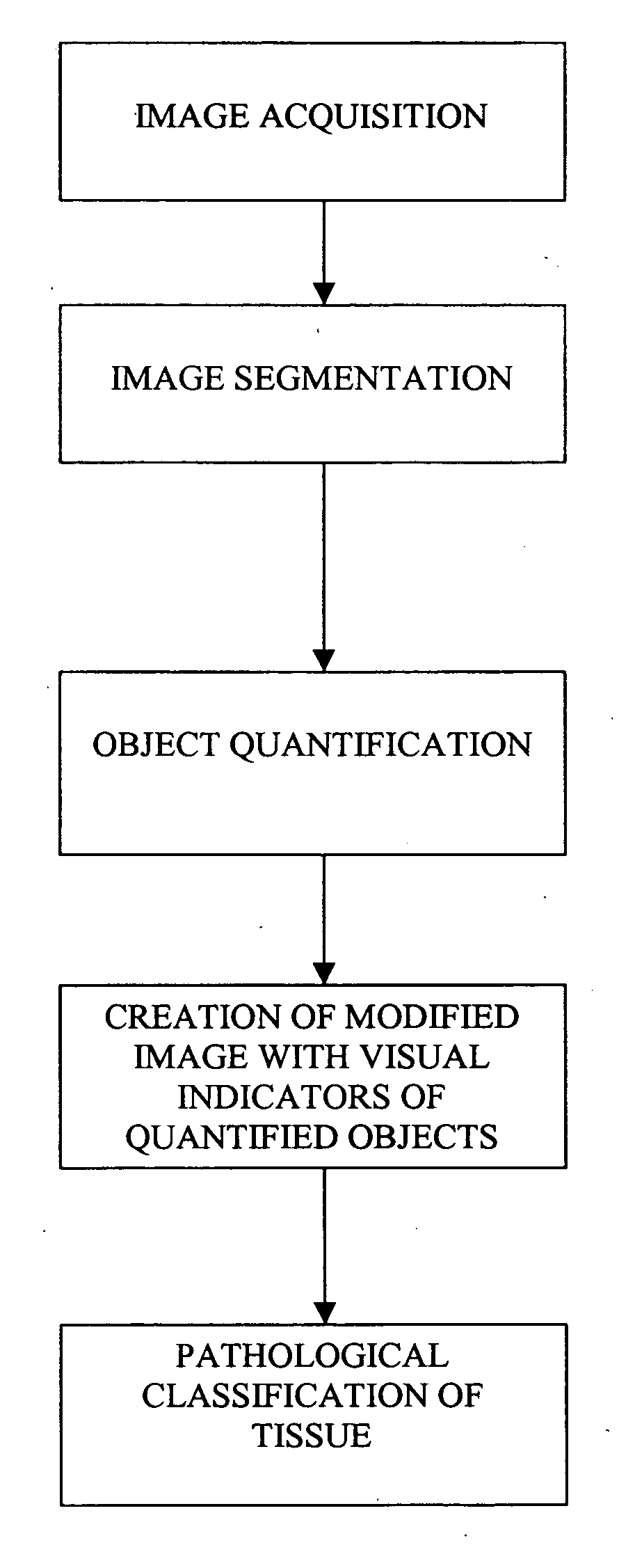
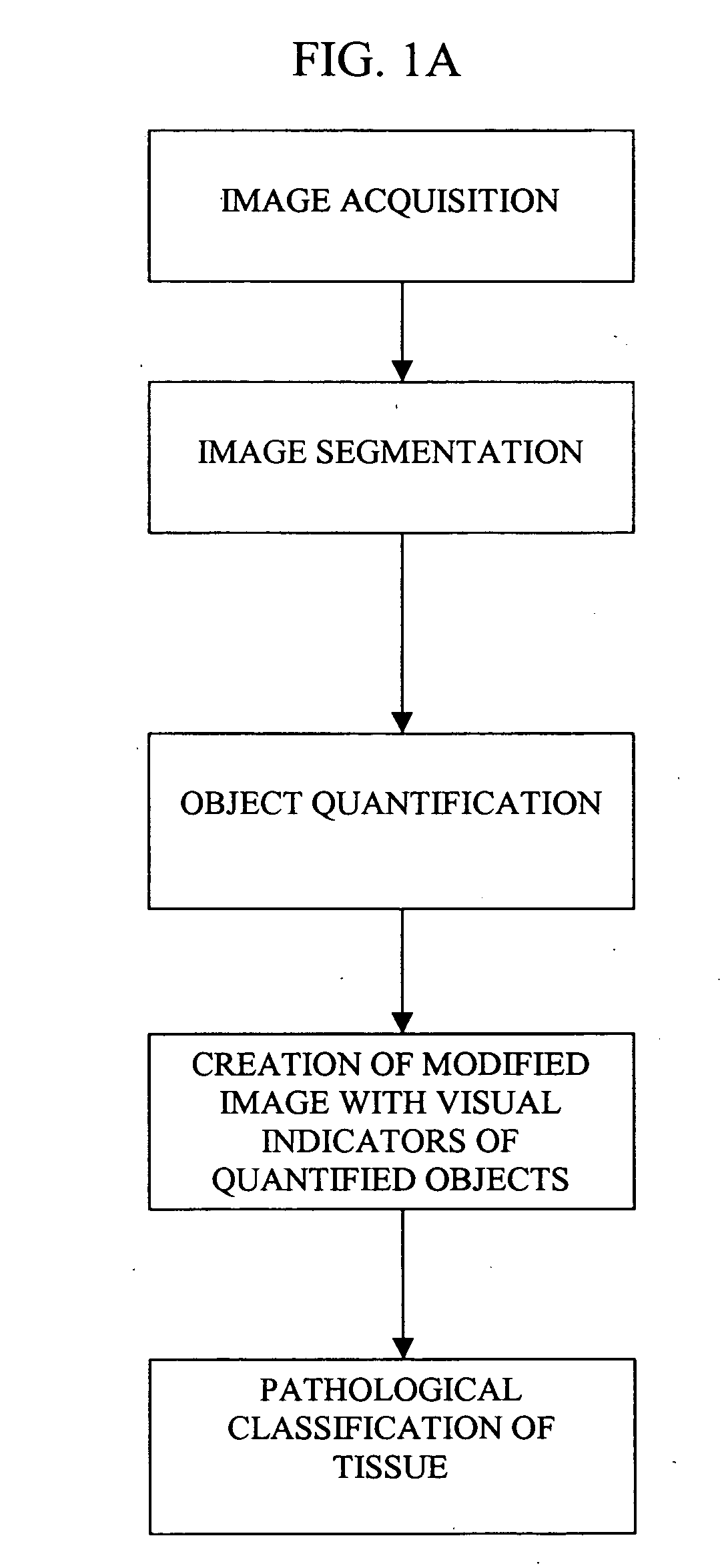
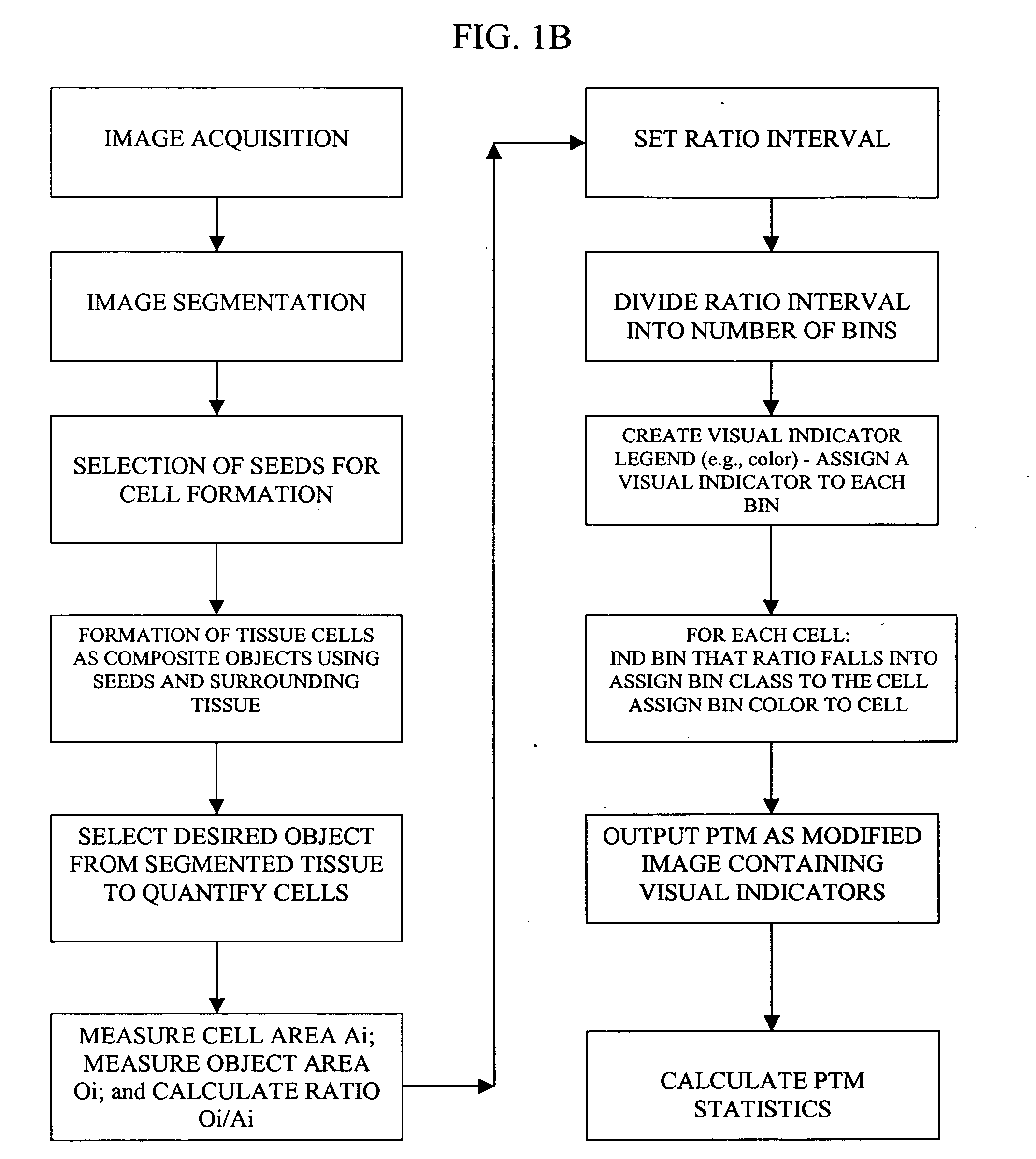
Pathological tissue mapping
a tissue mapping and pathology technology, applied in the field of molecular biology, histology, clinical diagnostics, can solve the problems of slow, difficult, prone to errors, and prior art methods and systems relating to the study of diseas
- Summary
- Abstract
- Description
- Claims
- Application Information
AI Technical Summary
Benefits of technology
Problems solved by technology
Method used
Image

Examples
example 1
Liver Tissue Image Segmentation—Portal Tract
[0084] Analysis of Bile Duct demonstrates that it is a tissue structure consisting of lumen (white area on the original image fragment FIG. 4A; colored yellow on the segmented image FIG. 4B) lined by simple cuboidal or columnar epithelium (epithelial nuclei painted by blue color on the segmented image).
Vessels
Hepatic Vein
[0085] Analysis of the Hepatic Vein (see FIG. 5A original image; FIG. 5B segmented image) which is the largest diameter vessel, reveals it to be another tissue structure consisting of lumen (large white area on the original image fragment colored light grey on the segmented image) which has the typical, thin-walled structure relative to the diameter of the lumen and irregular outline of all veins (colored aquamarine on the segmented image).
Hepatic Artery and Arterioles
[0086] The smaller diameter, thick-walled vessels with the typical structure of arterioles and arteries are branches of the Hepatic Arter...
example 2
[0092] This study was undertaken to demonstrate neural network and linear discriminant analysis (LDA) modeling capabilities of the present invention. Specifically, the study involved the acquisition and analysis of sections of rat liver with the overall objective being to classify the sections as normal or abnormal. Being able to automate this process while simultaneously achieving a high-level of classification accuracy allows for the creation of a high-throughput platform used to objectively screen for toxicities in pre-clinical studies.
[0093] The study was divided into two phases. The initial phase used a set of 100 rat liver sections as a training set; 80 normal liver sections and 20 abnormal. The image analysis process was then applied to an unlabeled set of 100 rat liver sections in the second phase of the study in which the statistical models designed in the training phase were tested.
[0094] Both the training and test set of rat liver sections were H&E-stained sl...
PUM
 Abstract
Description
Claims
Application Information