Partitioning method and device for endoscope fluorescence image and storage medium

A fluorescence image and image segmentation technology, applied in the field of medical molecular imaging, can solve the problems of complex anatomical structure and difficult medical image segmentation, and achieve the effect of overcoming non-homogeneity, improving computing speed, and improving training speed.

Active Publication Date: 2018-05-01
BEIJING DIGITAL PRECISION MEDICAL TECH CO LTD
View PDF7 Cites 19 Cited by
  • Summary
  • Abstract
  • Description
  • Claims
  • Application Information

AI Technical Summary

Problems solved by technology
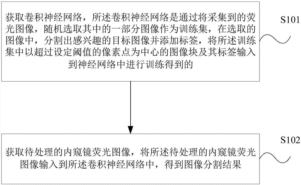
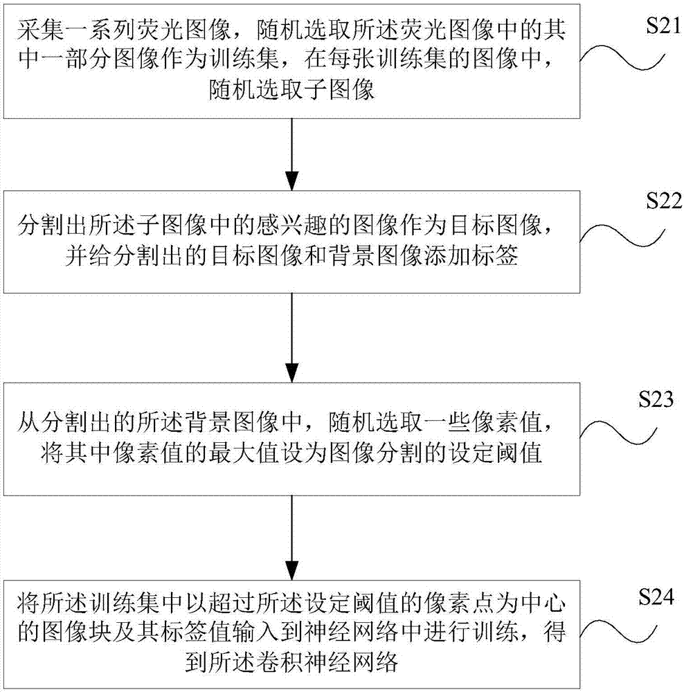
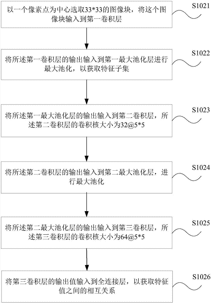
In addition, human anatomy is very complex and varies from person to

Method used


Image

  • Partitioning method and device for endoscope fluorescence image and storage medium
  • Partitioning method and device for endoscope fluorescence image and storage medium
  • Partitioning method and device for endoscope fluorescence image and storage medium

Examples


Embodiment Construction

[0020] The following will clearly and completely describe the technical solutions in the embodiments of the present invention with reference to the accompanying drawings in the embodiments of the present invention. Obviously, the described embodiments are only some, not all, embodiments of the present invention. Based on the embodiments of the present invention, all other embodiments obtained by persons of ordinary skill in the art without making creative efforts belong to the protection scope of the present invention.

[0021] Those skilled in the art know that the embodiments of the present invention can be implemented as a system, device, device, method or computer program product. Therefore, the present disclosure may be embodied in the form of complete hardware, complete software (including firmware, resident software, microcode, etc.), or a combination of hardware and software.

[0022] The principle and spirit of the present invention will be explained in detail below w...


PUM


Abstract

The invention provides a partitioning method and device for an endoscope fluorescence image and a storage medium. The method comprises the steps that a convolutional neural network is acquired, wherein the convolutional neural network is obtained in the mode that a part of image is randomly selected from a collected fluorescence image to serve as a training set, a target image of interest is partitioned from the selected image, a tag is added to the target image, and an image bock with a pixel point exceeding a set threshold being the center in the training set and a tag of the image block areinput into a neural network for training; and the to-be-processed endoscope fluorescence image is acquired and input into the convolutional neural network, and an image partitioning result is obtained. According to the method, the convolutional neural network is utilized to convert a partitioning problem of the optical operation navigation endoscope fluorescence image into a dichotomy problem ofimage pixel points; and through continuous training and optimization of a whole system, inhomogeneity of the optical operation navigation endoscope fluorescence image is effectively overcome, and thetarget of interest in the image is accurately extracted.

Description

technical field [0001] The invention relates to the field of medical molecular imaging, in particular to an endoscope fluorescence image segmentation method, device and storage medium. Background technique [0002] Medical image processing and analysis is an interdisciplinary subject that has emerged in recent years, making medical images gradually develop from film to digital. The traditional medical imaging technology only obtains the image data of a certain section of the human body, and then the doctor makes a diagnosis through the film or observes it on the monitor. However, what doctors can observe is only two-dimensional images, and they can only observe the images in a fixed way, and the obtained diagnosis results are subject to empirical judgment, which largely depends on the doctor's clinical experience. Through graphic image technology, the image data can be arbitrarily enlarged, reduced, rotated, three-dimensional reconstruction and other processing, so that doc...

Claims


Application Information

Patent Timeline
IPC IPC(8): G06T7/10G06T7/136G06N3/04G06N3/08
CPCG06T7/10G06T7/136G06T2207/10068G06T2207/20081G06T2207/20084
Inventor 迟崇巍王丽
Owner BEIJING DIGITAL PRECISION MEDICAL TECH CO LTD
Why Eureka
Try Eureka