A heptamethine fluorescent dye and its application in precise diagnosis and treatment of tumors
A fluorescent dye, tumor technology, applied in the field of near-infrared fluorescent dyes, can solve problems such as changing the specificity and affinity of target molecules
- Summary
- Abstract
- Description
- Claims
- Application Information
AI Technical Summary
Problems solved by technology
Method used
Image
Examples
Embodiment 1
[0043] The preparation of compound I of the present invention
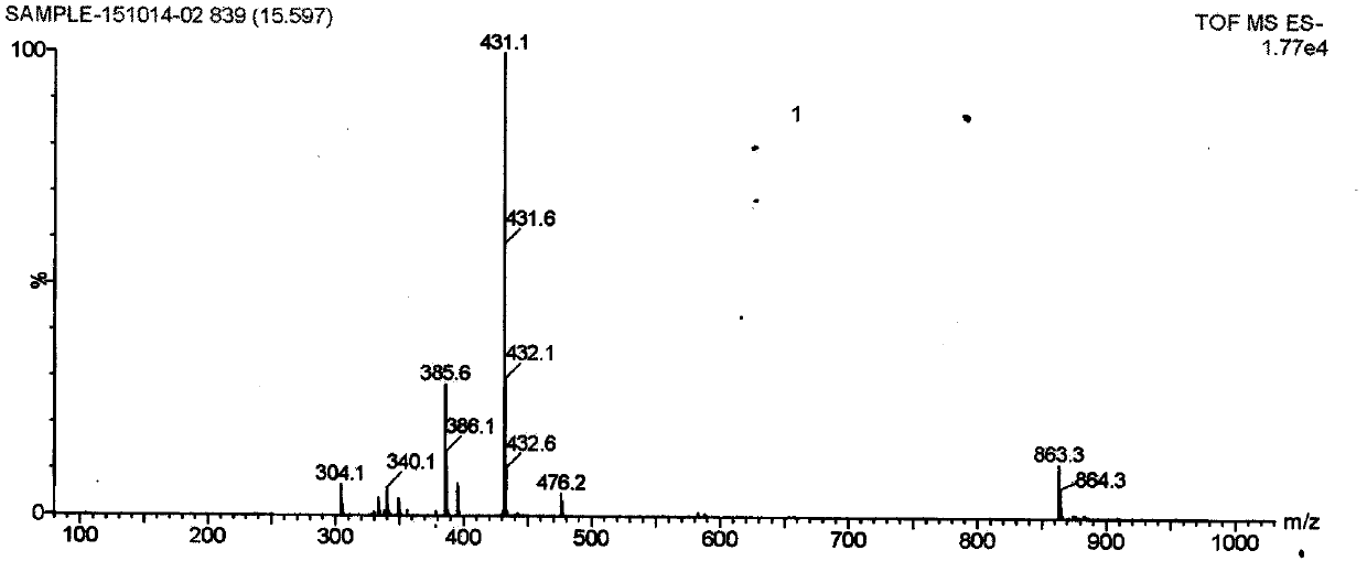
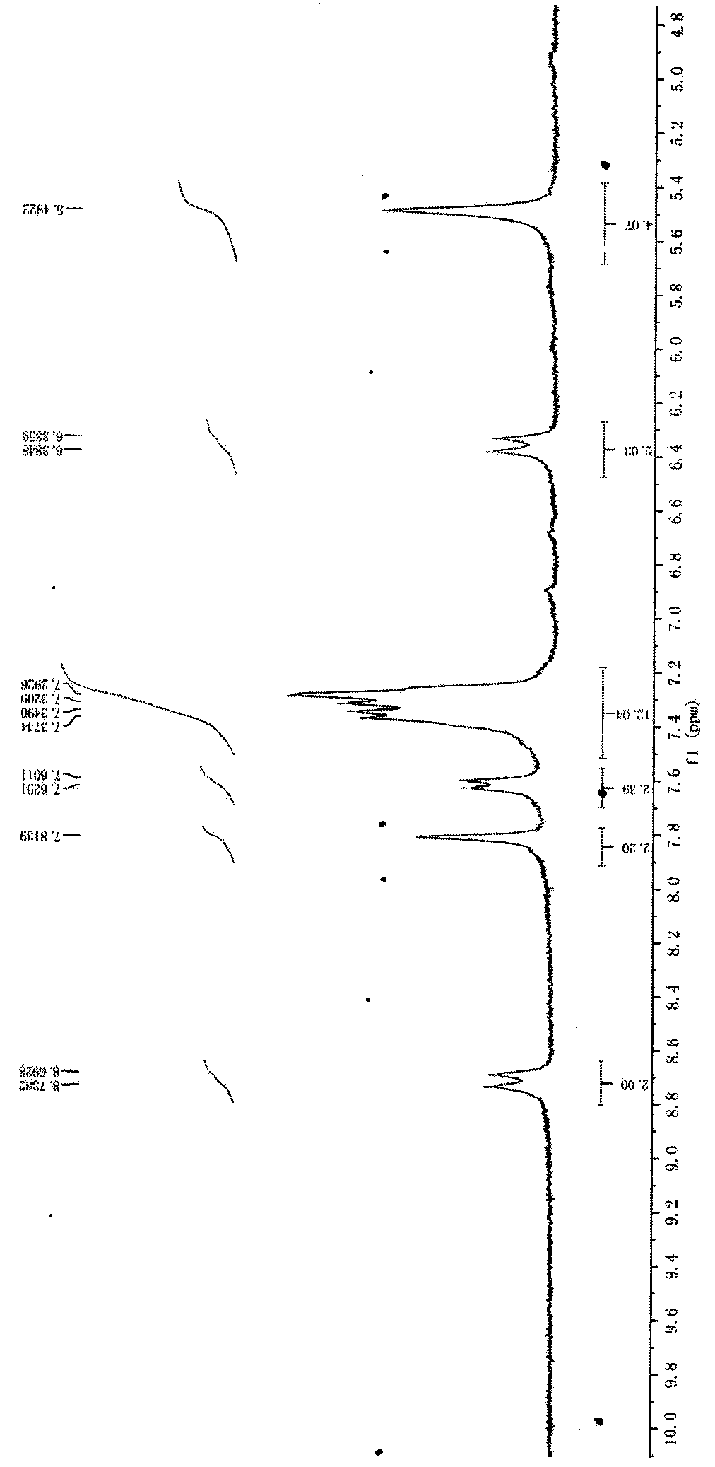
[0044] 1. Add phenylhydrazine p-sulfonic acid (5g) to methanol solution of methyl isopropyl ketone (8.55 / 15ml), heat the solution to 117°C, stir for 5h, and evaporate the solvent. Then 50 ml of diethyl ether was added to the oily product to obtain a pink powder, namely compound (3). Then, brown red powder (6g) is added in sodium hydroxide (1.5 grams) solution, solvent is methanol (10 milliliters) and isopropanol (10 milliliters), after this solution is stirred at 82 ℃ for 15 minutes, is cooled to room temperature , a large amount of No. (3) compound was separated and used for the next step of purification. m / z=261.27, NMR spectrum (500 MHz, chloroform) δ7.90 (d, 2H) 7.49 (s, H) 2.10 (s, 3H) 1.44 (s, 6H)
[0045] 2. Compound 3 (6 g) and 1-(bromomethyl)benzene (4.77 ml) were dissolved in 36 ml of toluene. The mixture was stirred and refluxed under nitrogen for 5 h, when the mixture was cooled, the solvent was rem...
Embodiment 2
[0050] Absorption of ICG-03 dye by different cell lines and human tumor-bearing nude mouse models
[0051] The logarithmic growth phase of U87 (glioma cells), MCF7 (breast cancer cells), HepG2 (human liver cancer cells), A549 (human lung cancer cells), MDA-MB-231 (human breast cancer cell lines), Panc1 (Human pancreatic cancer cell line) and L02 (human normal liver cells) were digested with trypsin, and the cells were transferred to laser confocal culture dishes, and the cell density was about 3×10 5 piece / cm 2 . Afterwards, the confocal culture dish was placed in a constant temperature cell culture incubator (37°C, 5% O 2 ) for 24 hours. After the cells adhered to the wall, 0.2 mL of ICG-03 solution filtered through a filter membrane was added and incubated for 2 hours. Cells were then washed twice with cold PBS solution (pH 7.4) for dual-channel fluorescence imaging by confocal laser microscopy.
[0052] Observing the fluorescence intensity of ICG-03 in tumor cells appa...
Embodiment 3
[0056] Compound Safety Test
[0057] In order to investigate the toxic and side effects of drugs on normal tissues, tissue sections and pathological studies are required. 30 rats aged 6 weeks, with a body weight of 200±10g, were intraperitoneally injected with ICG-03 dye 10mg / kg, and the rats were sacrificed four weeks later. Observe whether there are any changes in rat heart, liver, spleen, lung and kidney, fix the tissues in 10% formalin, and do HE staining on sections. There are no significant changes in rat heart, liver, spleen, lung and kidney, and histopathology is not significantly different from that of normal control rats. The dose is about 100 times the dose used in mouse imaging detection. The weight of the mouse is 20g. The dose is 0.1 mg / kg, which proves the safety of this dose. The results show that the dye has no toxicity to animals and cells, and shows good potential for clinical tumor detection and diagnosis.
PUM
| Property | Measurement | Unit |
|---|---|---|
| weight | aaaaa | aaaaa |
Abstract
Description
Claims
Application Information
 - R&D
- Intellectual Property
- Life Sciences
- Materials
- Tech Scout
- Unparalleled Data Quality
- Higher Quality Content
- 60% Fewer Hallucinations
Browse by: Latest US Patents, China's latest patents, Technical Efficacy Thesaurus, Application Domain, Technology Topic, Popular Technical Reports.
© 2025 PatSnap. All rights reserved.Legal|Privacy policy|Modern Slavery Act Transparency Statement|Sitemap|About US| Contact US: help@patsnap.com


