
Automated imaging method and system

An imaging method and automatic technology, which is applied in the fields of medical automatic diagnosis, image enhancement, image analysis, etc., can solve the problems of inaccurate diagnosis and time-consuming, and achieve the effect of reducing operation time, reducing difficulty, and improving accuracy

Active Publication Date: 2021-12-07
SHANGHAI UNITED IMAGING HEALTHCARE
View PDF10 Cites 0 Cited by
  • Summary
  • Abstract
  • Description
  • Claims
  • Application Information

AI Technical Summary

Problems solved by technology

[0003] Aiming at the above-mentioned problems that manual operation is not only time-consuming but also may lead to inaccurate diagnosis, the purpose of the present invention is to provide an automatic imaging method and system, which can improve the efficiency and accuracy of diagnosis and reduce the difficulty of diagnosis

Method used


Image

  • Automated imaging method and system
  • Automated imaging method and system
  • Automated imaging method and system

Examples


Embodiment Construction

[0035] In order to more clearly illustrate the technical solutions of the embodiments of the present application, the following briefly introduces the drawings that need to be used in the description of the embodiments. Obviously, the accompanying drawings in the following description are only some examples or embodiments of the present application, and those skilled in the art can also apply the present application to other similar scenarios. It should be understood that these exemplary embodiments are given only to enable those skilled in the relevant art to better understand and implement the present invention, but not to limit the scope of the present invention in any way. Unless otherwise apparent from context or otherwise indicated, like reference numerals in the figures represent like structures or operations.

[0036] It should be understood that the terms "system", "unit", "module" and / or "engine" described above and below are used to distinguish different levels of ...


PUM


Abstract

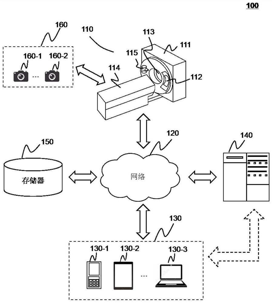
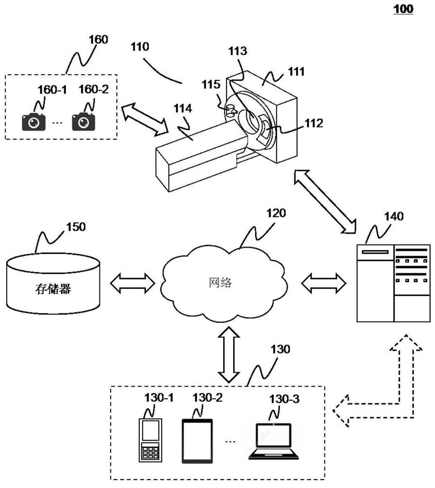
The invention discloses an automatic imaging method and system. The method includes: acquiring image data of a target object placed on a scanning bed; determining first position information of the target object by a processor based on the image data; determining by the processor based on the image data second location information related to a scanning area of ​​the target object; and scanning the target object by an imaging device based on the first location information and the second location information. The automatic imaging method reduces the user's operation time before clinical scanning, and can also avoid the user's mistakes in manual operation, thereby improving the efficiency and accuracy of clinical examination.

Description

technical field [0001] The present application relates to an imaging method and system, in particular to an automatic imaging method and system. Background technique [0002] Medical imaging often requires the ability to accurately and repeatably position patients during diagnostic scans and treatments. During scanning, the scanning table supporting the patient needs to be moved into position. Typically, the scanning table can be moved manually by the operator. At the same time, the scanner parameters also need to be manually set by the operator, so that the patient can be scanned in multiple different positions. These manual operations are time-consuming and can lead to inaccurate diagnoses. Therefore, an effective automatic imaging mechanism is needed to improve scanning efficiency and diagnostic accuracy. Contents of the invention [0003] In view of the above-mentioned problems that the manual operation is not only time-consuming but also may lead to inaccurate dia...

Claims


Application Information

Patent Timeline
Patent Type & Authority Patents(China)
IPC IPC(8): G06K9/00G06K9/20
CPCG06V40/10G06V10/10G06V10/22A61B6/032A61B6/037A61B6/0407A61B6/5205A61B6/563G06T2207/10081G06T2207/20081G06T2207/20084G06T7/70G16H30/20G16H50/50G16H50/20G16H40/63A61B6/545
Inventor 全国涛
Owner SHANGHAI UNITED IMAGING HEALTHCARE
Learn More