
Method for building three-dimensional tooth-and-jaw fusion model
A technology that integrates models and jaws, applied in teaching models, dentistry, articulators, etc., can solve problems such as affecting clinical results, soft tissue data distortion, errors, etc., to save clinical work time, manpower and material resources, and reduce CT image errors. , the effect of reducing the number of registrations
- Summary
- Abstract
- Description
- Claims
- Application Information
AI Technical Summary
Problems solved by technology
Method used
Image

Examples
Embodiment Construction
[0035] The present invention is further described below in conjunction with accompanying drawing.
[0036] A method for establishing a three-dimensional dento-jaw fusion model, comprising the following steps:
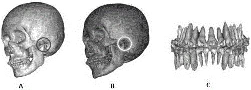
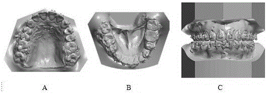
[0037] 1) Data acquisition, put the ironed and cut horseshoe-shaped wax sheet into the patient's mouth, bite, that is, use the wax sheet to record the patient's occlusal position, which is the common and unique occlusal position of the image data and model data; relax the facial soft tissue , take head CT in the above occlusal state, and complete the collection of image data; perform conventional intraoral silicone rubber impression material on the patient to make an impression, perfuse the plaster model, and use the above wax slice to determine the occlusal relationship of the plaster model to complete the collection of model data;
[0038] 2) Image data processing, import the cranial CT file described in step 1) into Mimics, reconstruct the three-dimensional images of...
PUM
 Abstract
Description
Claims
Application Information