Iterative Fundus Image Vessel Segmentation Method Based on Range Modulation Loss

A fundus image and blood vessel technology, applied in the field of image processing, can solve the problems of vascular structure constraints, models that cannot be learned to improve segmentation results, etc., and achieve the effect of improving accuracy and robustness

Active Publication Date: 2020-07-31
SHANGHAI JIAOTONG UNIV
View PDF3 Cites 0 Cited by
  • Summary
  • Abstract
  • Description
  • Claims
  • Application Information

AI Technical Summary

Problems solved by technology

However, the existing deep learning methods simply transform the fundus image blood vessel segmentation problem into a binary classification problem without further constraints on the blood vessel structure. At the same time, if the network output results are not ideal, the learned model cannot be further utilized. Improve Segmentation Results

Method used


Image

  • Iterative Fundus Image Vessel Segmentation Method Based on Range Modulation Loss
  • Iterative Fundus Image Vessel Segmentation Method Based on Range Modulation Loss
  • Iterative Fundus Image Vessel Segmentation Method Based on Range Modulation Loss

Examples


Embodiment

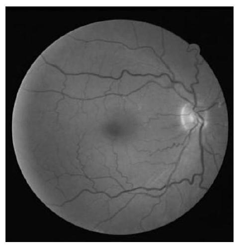
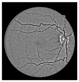
[0074] The fundus image used in this embodiment comes from a public data set, and the hardware device used is a GPU workstation, including Intel Xeon CPU E5-2620 and GeForce GTX 1080GPU, as shown in Figure 2 (a) is the G channel component of the training image.

[0075] (1) Normalize the original image (ie fundus image)

[0076] Since fundus images from different sources have inconsistent image resolution, brightness, and contrast, it is necessary to normalize before inputting the segmentation model (ie dense convolutional neural network model) to ensure the stability of the segmentation algorithm. The specific implementation steps are as follows:

[0077] 1) Unify the diameter of the field of view, that is, take the position of the middle 1 / 2 height of the original image to estimate the size of the field of view of the original image, sum the values ​​of the RGB channels of each pixel along the width direction of the original image, and then Binarize each pixel:

[0078] B ...


PUM


Abstract

The invention discloses an iterative eye fundus image blood vessel segmentation method based on the distance modulation loss, which comprises the steps of (0) collecting a color eye fundus image to form an original image; (1) performing normalization processing on the original image; (2) iteratively training a dense convolution neural network based on the distance modulation loss; and (3) carryingout iterative blood vessel segmentation by using the trained dense convolution neural network. The method can process color eye fundus images under different collection conditions, can provide interactive blood vessel segmentation experience for ophthalmologists, is more robust for blood vessel detection and provides a reliable guarantee for subsequent auxiliary diagnosis.

Description

technical field [0001] The invention belongs to the field of image processing, and in particular relates to an iterative fundus image blood vessel segmentation method based on distance modulation loss. Background technique [0002] The automatic fundus image blood vessel segmentation technology with interactive mode can provide ophthalmologists with fast service for subsequent measurement of intraretinal blood vessels, and help doctors efficiently evaluate potential diabetes, hypertension, arteriosclerosis and other diseases. At present, there are a large number of algorithms and technologies for blood vessel segmentation in fundus images at home and abroad, mainly including two categories of technologies: segmentation methods with unsupervised training and segmentation methods with supervised training. [0003] The most representative segmentation method based on unsupervised training is the Matched Filters method. This method obtains the grayscale feature of the blood vess...

Claims


Application Information

Patent Timeline
Patent Type & Authority Patents(China)
IPC IPC(8): G06T7/11
CPCG06T7/11G06T2207/10024G06T2207/20081G06T2207/30041
Inventor 杨杰周磊
Owner SHANGHAI JIAOTONG UNIV
Why Eureka
Try Eureka