
Method for extracting center line of coronary artery blood vessel

A coronary artery and centerline technology, applied in the field of medical blood vessel image processing, can solve the problems of error-prone centerline extraction, poor over-effect at bifurcation points, inconsistent doctor's operation, etc., so as to improve diagnostic efficiency, ensure accuracy, Guaranteed uniformity

Active Publication Date: 2018-06-08
SUZHOU RAINMED MEDICAL TECH CO LTD
View PDF3 Cites 31 Cited by
  • Summary
  • Abstract
  • Description
  • Claims
  • Application Information

AI Technical Summary

Problems solved by technology

The disadvantages are: the iterative calculation process is time-consuming, and it is easily affected by the image quality, and the transition effect at the bifurcation point is poor
The disadvantages are: this method is based on the local method, and the global information is not considered in the tracking process, so sometimes the obtained centerline will be incomplete. Large or branched locations, centerline extraction is prone to errors
The disadvantage is that the generated centerline is not accurately located in the center of the object, but tends to be close to the inner wall of the object, so it often requires later adjustments, and manual interaction is required to specify the start point and end point
This method is more popular, but the disadvantage is that the start and end points of the centerline need to be specified
[0007] All of the above methods need to specify the start point and end point through interaction before generating the centerline. All blood vessels cannot be fully identified automatically, and the operations of each doctor are not uniform, resulting in diversity of results.

Method used


Image

  • Method for extracting center line of coronary artery blood vessel
  • Method for extracting center line of coronary artery blood vessel
  • Method for extracting center line of coronary artery blood vessel

Examples


Embodiment

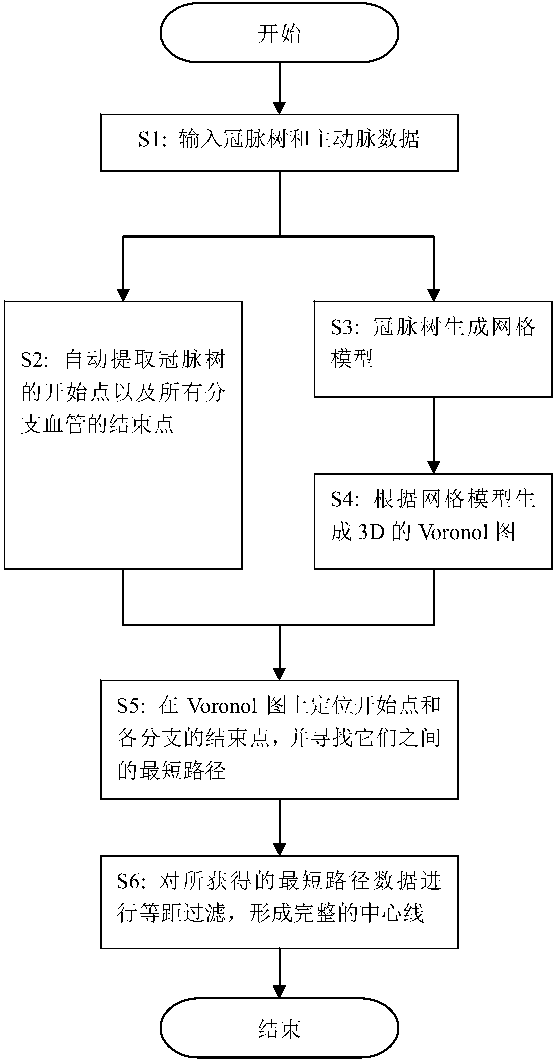
[0039] Example: refer to Figures 1 to 3 As shown, a method for extracting the centerline of the coronary artery comprises the following steps:

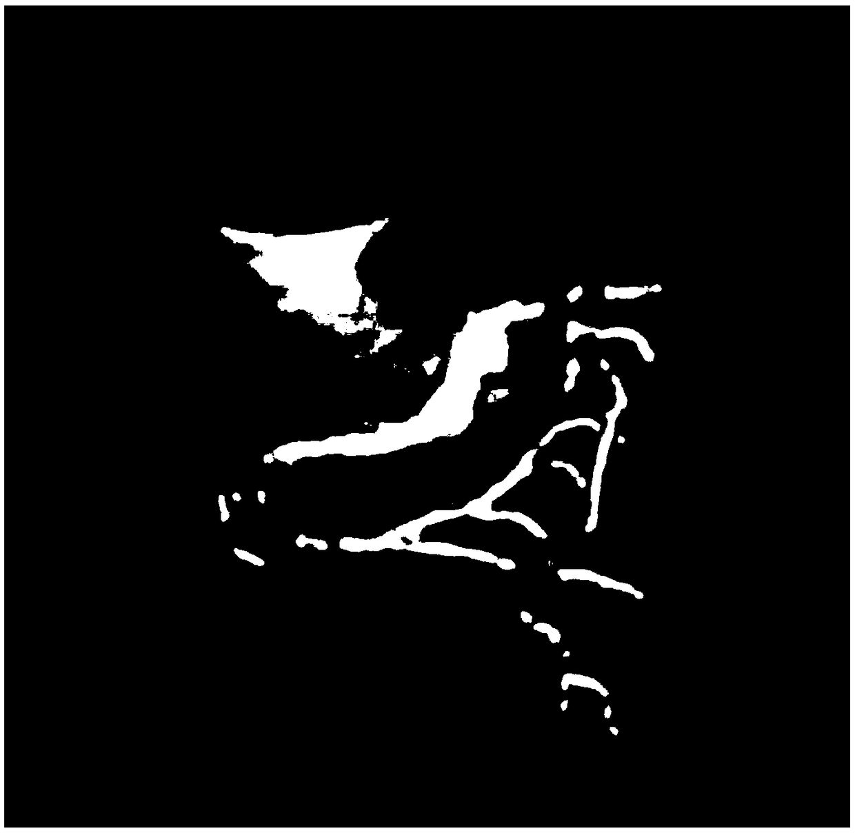
[0040] Step S1: input the data of coronary tree and aorta;

[0041] Step S2: According to the coronary tree and aorta data in step S1, the intersection part is obtained from the overlapping part of the data, and its centroid M is obtained. The three-dimensional coordinate formula of the centroid M is:

[0042]

[0043] where m i is the coordinate point (x i ,y i ,z i ) at the gray value;

[0044] Then, with the centroid M as the starting point, the coronary tree data is gradually traversed to the end of each branch vessel in the way of nibbling, and the end points of all branch vessels are obtained. The specific method for obtaining the end points of all branch vessels is as follows:

[0045] Step A: Set the list List to represent the collection of end points of all branch vessels, the list ListA to represent the center of t...


PUM


Abstract

The invention discloses a method for extracting a center line of a coronary artery blood vessel. The method comprises the steps that S1, coronary artery tree data and aorta data are input; S2, a starting point of a coronary artery tree and ending points of all branch blood vessels are automatically extracted; S3, a Marching Cubes algorithm is used to make the coronary artery tree data into grid model data; S4, Delaunay triangulation processing is performed on all points on a grid, and a three-dimensional Voronoi diagram is generated; S5, the starting point and the ending points of all the branch blood vessels are located on the Voronoi diagram, and the shortest path between the starting point and the ending points is calculated to obtain several corresponding curves; and S6, isometric filtering is performed on the obtained curves, and complete center line data is formed. The method has the advantages that the starting point and the ending points of the coronary artery blood vessel areautomatically acquired, and the accuracy of the center line is guaranteed; and the powerful processing capability of a computer is fully utilized, the step of acquiring the starting point and the ending points through a manual interaction mode is omitted, and the efficiency of doctor diagnosis is greatly improved.

Description

technical field [0001] The invention belongs to the field of medical blood vessel image processing, and in particular relates to a method for extracting the center line of a coronary artery. Background technique [0002] With the development and progress of modern science and technology, the application of medical imaging technology is becoming more and more extensive, and it is favored by more and more doctors and accepted by more and more patients. Contrast images are often used by doctors as a reference for the diagnosis of clinical diseases and formulation of treatment plans. Due to the influence and constraints of doctors' subjective factors, it is not enough to analyze and judge the contrast images only by doctors' clinical experience and vision. It has become an inevitable trend to use the efficiency and accuracy of computers to quantitatively analyze contrast images. In clinical practice, doctors hope to obtain quantitative analysis results by analyzing blood vessel...

Claims


Application Information

Patent Timeline
IPC IPC(8): G06T7/00G06T7/187G06T7/62G06T7/66
CPCG06T7/0012G06T2207/20156G06T2207/30101G06T2210/41G06T7/187G06T7/62G06T7/66
Inventor 霍云飞冯亮吴星云霍勇龚艳君李建平易铁慈杨帆
Owner SUZHOU RAINMED MEDICAL TECH CO LTD
Features
  • Generate Ideas
  • Intellectual Property
  • Life Sciences
  • Materials
  • Tech Scout
  • Unparalleled Data Quality
  • Higher Quality Content
  • 60% Fewer Hallucinations
Learn More