Intra-operative pathological tissue quick extracting device

An extraction device and tissue technology, applied in the field of intraoperative pathological tissue rapid extraction devices, can solve problems such as affecting the diagnostic accuracy, squeezing the front end of the syringe or even the needle, and air squeezing into the human body, achieving ingenious structural design and ensuring safety. The effect of increasing the friction force

Active Publication Date: 2018-06-22
关慧娟
View PDF11 Cites 3 Cited by
  • Summary
  • Abstract
  • Description
  • Claims
  • Application Information

AI Technical Summary

Problems solved by technology

However, there are still many deficiencies in the use of the tissue extractor in this case, for example: (1) After the tissue extraction is completed, the extraction cavity needs to be pulled out gradually from the rear end of the needle, and the rear end of the syringe is not sealed, which is prone to bacteria , there is a risk of bacteria and viruses adhering to the tissue in the extraction cavity during the process of pulling out the extraction cavity, which will affect the accuracy of diagnosis; (2) During the process of extracting tissue, the stopper connected with the extraction cavity moves to the front end of the piston inner cavity , the air originally accumulated at the front end of the stopper is squeezed into the front end of the syringe or even the needle, and there is a risk of air being squeezed into the human body when the design is unreasonable; (3) The process of gradually pulling the extraction chamber from the rear end of the needle delays pathology detection time, so that the extraction speed of pathological tissue still cannot meet the purpose of rapid tissue extraction

Method used


Image

  • Intra-operative pathological tissue quick extracting device
  • Intra-operative pathological tissue quick extracting device
  • Intra-operative pathological tissue quick extracting device

Examples


Embodiment 1

[0028] Example 1 A rapid extraction device for intraoperative pathological tissue

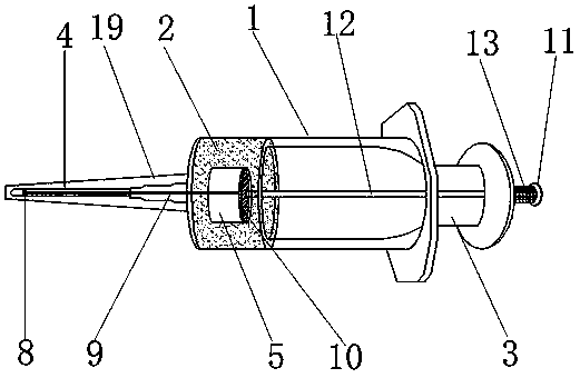
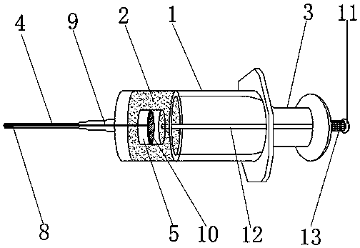
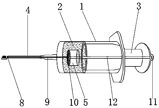
[0029] like Figure 1-6 In the shown embodiment, a device for rapidly extracting intraoperative pathological tissue includes a syringe 1, a piston 2 disposed in the syringe, a hollow push-pull rod 3 fixedly connected to the end of the piston 2, and a needle 4 disposed at the front end of the syringe 1 , the inner front end of the needle 4 is provided with an "I"-shaped tissue pushing mechanism extending to the inside of the piston 2; the rear end of the hollow push-pull rod 3 is provided with a "T"-shaped pushing mechanism, and the "T"-shaped pushing mechanism is The rear end of the hollow push-pull rod 3 penetrates the interior of the hollow push-pull rod 3 and extends to the rear end of the piston 2; a cavity 5 is provided inside the piston 2, and a first through hole 6 communicating with the cavity 5 is provided at the front end of the piston 2 , The rear end of the piston 2 is provided wit...

Embodiment 2

[0034] Example 2 A rapid extraction device for intraoperative pathological tissue

[0035] The structure of this embodiment is basically the same as that of Embodiment 1, the only difference being that: the front end surface of the front push plate 8 in this embodiment is provided with a slice 16 extending to the opening of the needle point of the needle head 4, and the slice 16 is as follows: Figure 7 As shown, it is a horizontal slice, and the horizontal slice is used to cut the pathological tissue when extracting the pathological tissue, so that the cell layers are more distinct, and two cell smears are prepared at a time for comparative observation.

Embodiment 3

[0036] Example 3 A rapid extraction device for intraoperative pathological tissue

[0037] The structure of this embodiment is basically the same as that of Embodiment 1, the only difference being that: the front end surface of the front push plate 8 in this embodiment is provided with a slice 16 extending to the opening of the needle point of the needle head 4, and the slice 16 is as follows: Figure 8 As shown, it is a "ten" type slice. "Ten"-shaped slices are used to cut pathological tissues when extracting pathological tissues, so that the cell layers are more distinct, and four cell smears are prepared at a time for comparative observation.


PUM


Abstract

The invention relates to a pathology determining auxiliary device, in particular to an intra-operative pathological tissue quick extracting device which comprises a syringe, a piston, a hollow push-pull rod and a needle. The piston is arranged in the syringe, the hollow push-pull rod is fixedly connected with the tail end of the piston, the needle is arranged at the front end of the syringe, an 'H'-shaped tissue pushing mechanism which extends into the piston is arranged at the front end of the inside of the needle, and a 'T'-shaped pushing mechanism is arranged at the rear end of the hollow push-pull rod, penetrates into the hollow push-pull rod from the rear end of the hollow push-pull rod and extends to the rear end of the piston; a cavity is arranged inside the piston, a first throughhole which is communicated with the cavity is formed in the front end of the piston, and a second through hole which is communicated with the cavity is formed in the rear end of the piston. The intra-operative pathological tissue quick extracting device has the advantages that tissues can be extracted by the intra-operative pathological tissue quick extracting device in sterile environments, a seal cover can sleeve the intra-operative pathological tissue quick extracting device after the intra-operative pathological tissue quick extracting device is disinfected and can be directly pulled downin operation, and accordingly the intra-operative pathological tissue quick extracting device can be used; tissue extracting procedures are convenient and speedy, and the intra-operative pathologicaltissue quick extracting device is high in safety.

Description

technical field [0001] The invention relates to a device for assisting in determining pathology, in particular to a device for rapidly extracting pathological tissue during operation. Background technique [0002] Tumor pathological examination is one of the most important methods for tumor diagnosis. Pathological examination can determine the diagnosis, tissue source, nature and extent of the tumor, and provide an important basis for clinical treatment. The accuracy of pathological examination results during the operation and the control of the operation time are crucial to the safety of the patient. Therefore, the method of pathological determination during the operation must be efficient and accurate. Traditional pathological film production requires more than ten steps such as fixation, dehydration, and transparency. The whole process lasts 16-22 hours, resulting in 3-5 working days for routine pathological diagnosis. Due to the long time, it is only suitable for preope...

Claims


Application Information

Patent Timeline
Patent Type & Authority Applications(China)
IPC IPC(8): A61B10/02
CPCA61B10/0275A61B10/0283
Inventor 朱路英高峰宫福超李向阳
Owner 关慧娟
Why Eureka
Try Eureka