Method for determining tissue properties of tumors

A technology of tissue characteristics and measurement data, applied in the direction of radiodiagnostic instruments, applications, clinical applications of radiodiagnosis, etc., can solve the problems of limited additional information and unreliable clinical applications

Active Publication Date: 2018-06-29
SIEMENS HEATHCARE GMBH
View PDF11 Cites 0 Cited by
  • Summary
  • Abstract
  • Description
  • Claims
  • Application Information

AI Technical Summary

Problems solved by technology

However, in both methods the additional information obtained by evaluating CT images is limited and the method is not reliable enough for clinical application

Method used


Image

  • Method for determining tissue properties of tumors
  • Method for determining tissue properties of tumors
  • Method for determining tissue properties of tumors

Examples


Embodiment Construction

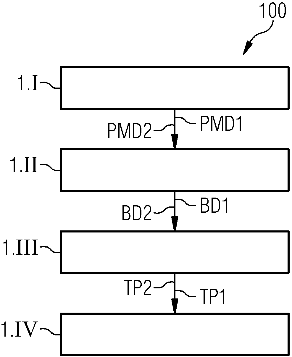
[0054] figure 1 A flowchart 100 is shown illustrating a method for determining tissue properties in an examination region. The patient is injected with a contrast medium in advance (ie before the procedure begins), which is delivered via the blood circulation to the examination area within the patient's body. Subsequently, in step 1.I, the examination region is irradiated with X-rays and two projection measurement data sets PMD1 , PMD2 assigned to different X-ray energy spectra, also called spectral projection measurement data, are acquired from the examination region. During the acquisition of spectral projection measurement data PMD1 , PMD2 in the examination region, previously injected contrast medium is present in the examination region. Then in step 1.II the image data BD1, BD2 are reconstructed on the basis of the acquired projection measurement data PMD1, PMD2. exist figure 1 In the exemplary embodiment shown, two pseudo-monoenergetic image data sets BD1 , BD2 are re...


PUM


Abstract

A method for determining tissue properties of tumors. The method includes acquiring contrast medium-enhanced projection measurement data (PMD1, PMD2 and PMD3) from the examination region, including atleast two spectral projection measurement data sets (PMD1, PMD2). Further, image data (BD1, BD2, BD3) is reconstructed based upon the acquired projection measurement data (PMD1, PMD2 and PMD3), the image data (BD1, BD2, BD3) including at least two spectral image data sets (BD1, BD2, BD3). Subsequently, texture parameters (TP1, TP2 and TP3) are determined based upon the reconstructed image data (BD1, BD2, BD3) and a parameter analysis is carried out based upon the parameter database. In addition, an image analysis apparatus (20) and a computed tomography system (40) are described.

Description

technical field [0001] The invention relates to a method for determining tissue properties in an examination region. The invention also relates to an image analysis device. Furthermore, the invention relates to a computer tomography system. Background technique [0002] Two-dimensional or three-dimensional image data are usually created with the aid of modern imaging methods, which can be used to visualize the imaged examination object and also for other purposes. [0003] Imaging methods are generally based on the detection of X-ray radiation, in which so-called "projection measurement data" are generated. For example, projection measurement data may be acquired by means of a computed tomography (CT) system. In a CT system, the combination of the X-ray source and the X-ray detector is arranged on a rotating gantry, and the X-ray detector and the X-ray source are installed opposite to each other. The combination usually rotates around the scanning space, and the object to...

Claims


Application Information

Patent Timeline
IPC IPC(8): A61B6/03A61B6/00G06T7/00G06T7/44
CPCG06T7/44A61B6/032A61B6/481A61B6/50A61B6/5217G06T7/0012G06T2207/10081G06T2207/30096A61B6/482A61B6/5235G06T2207/10144G06T2207/30004G06T2211/408G06T7/40G06T11/005G16H50/30G06T7/0016
Inventor T·弗洛尔B·施密特
Owner SIEMENS HEATHCARE GMBH
Why Eureka
Try Eureka