Supercharge Your Innovation With Domain-Expert AI Agents!

Tongue palate separation device when oral CT is shot

A separation device and oral cavity technology, applied in patient positioning for diagnosis, computerized tomography scanner, echo tomography, etc., can solve the problem of insufficient image quality, large deformation, poor separation effect of palate and tongue, etc. problems, to achieve the effect of regular shape, small deformation, and convenient clinical diagnosis

Pending Publication Date: 2018-07-17
SICHUAN UNIV
View PDF9 Cites 3 Cited by
  • Summary
  • Abstract
  • Description
  • Claims
  • Application Information

AI Technical Summary

Problems solved by technology

[0004] In recent years, individual radiology technicians have tried to let patients bite cotton rolls or gauze balls to separate the palate and tongue. Because cotton rolls or gauze balls are soft in texture, they deform greatly when biting, and the palate and tongue are separated. Poor separation and poor image quality

Method used


Image

  • Tongue palate separation device when oral CT is shot
  • Tongue palate separation device when oral CT is shot
  • Tongue palate separation device when oral CT is shot

Examples


Embodiment 1

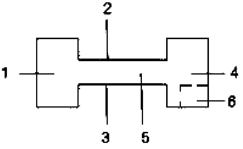
[0030] The tongue and palate separation device when shooting oral CT includes two occlusal pads and two tongue depressors. The palatal surface is connected as a whole; the separation pad is provided with the upper tooth surface and the lower tooth surface, which is used to place the upper and lower posterior teeth during shooting; the tongue depressor is vertically fixedly connected with the palatal surface of the tongue, and is used to fix the tongue during shooting to prevent the tongue improvement. The width of the upper and lower teeth is 1.2cm.

[0031] When in use, the back teeth on both sides bite the cushion at the same time. According to the size of the patient's mouth, two tongue depressors are used to fix the tongue.

[0032] The concave design in the middle of the occlusal pad is used for the occlusal fixation of the upper and lower posterior teeth. The occlusal pad is made of soft plastic, which has a certain degree of tolerance and deformation, so that teeth of...

Embodiment 2

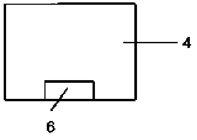
[0036] The tongue and palate separation device for taking oral CT includes two occlusal pads and a tongue depressor. The lingual and palatal surfaces are connected as a whole; the separation pad is provided with upper and lower tooth surfaces, which are used to place the upper and lower posterior teeth during shooting; the tongue depressor is vertically fixedly connected with the lingual and palatal surface, and is used to fix the tongue body during shooting to prevent tongue body elevation.

[0037] The tongue and palate surface is provided with a card slot, which is located at the bottom of the tongue and palate surface; the spatula is provided with a plug and a plate body, and the spatula and the tongue and palate surface are fixedly connected by inserting connection; when in use, the plug is inserted into the card slot, and the The spatula is fixedly connected with the palatal surface to form a whole.

[0038] In terms of size, the plug

Embodiment 3

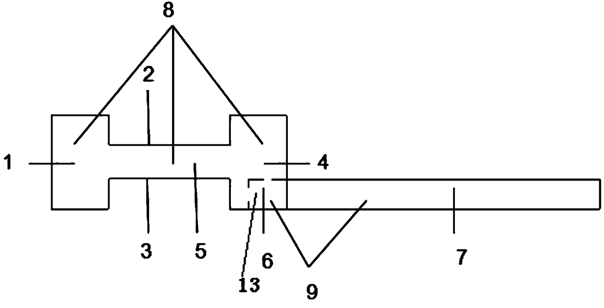
[0045]The tongue and palate separation device for taking oral CT includes two occlusal pads and a tongue depressor. The lingual and palatal surfaces are connected as a whole; the separation pad is provided with upper and lower tooth surfaces, which are used to place the upper and lower posterior teeth during shooting; the tongue depressor is vertically fixedly connected with the lingual and palatal surface, and is used to fix the tongue body during shooting to prevent tongue body improvement.

[0046] There is a card slot on the tongue and palate surface, which is located at the bottom of the tongue and palate surface. The tongue depressor and the tongue and palate surface are fixedly connected by inserting connection; the tongue depressor is equipped with a plug and a plate body. The plate is fixedly connected with the lingual-palatal surface to form a whole.

[0047] In terms of size, the plug

[0048] The dimensions of the buccal and lingual-palatal surfaces a...


PUM


Abstract

The invention belongs to the field of medical instruments, in particular to a tongue palate separation device when the oral CT is shot. The device is characterized in that two occlusal pads are arranged, the occlusal pads are H-shaped, a buccal side surface, a tongue palate surface and a separation pad are arranged, and the separation pad integrates the buccal side surface and the tongue palate surface together; the separation pad is provided with an upper tooth surface and a lower tooth surface for placing upper and lower back teeth when photographing is conducted; a spatula is fixedly connected with the tongue palate surface vertically and used for fixing a tongue body during shooting to prevent the tongue body from rising. The device is applied to CT or CBCT examination of goitre of tongue and palate, and the tongue and the palate are separated, thereby ensuring that the range of lesion can be clearly displayed during the shooting.

Description

technical field [0001] The invention belongs to the field of medical equipment, in particular to a tongue and palate separation device for shooting oral CT. Background technique [0002] In the narrow space of the oral cavity, the palate and tongue are anatomically close to each other. When the patient is in a resting state when taking oral CT, the lips are gently closed, the upper and lower teeth are slightly separated, and the surface of the palate is attached to the upper edge of the tongue. In the oral CT image taken at this time, the doctor cannot distinguish the soft tissue of the palate from the upper edge of the tongue. The tongue tissue is separated. [0003] When a lesion occurs in the palate or tongue of a patient, it is often necessary to take a CT scan of the oral cavity to judge the shape, scope and nature of the lesion. In clinical work, some radiologic technicians will instruct patients to keep their mouths open and tongues pressed in order to separate the ...

Claims


Application Information

Patent Timeline
IPC IPC(8): A61B6/04A61B6/03
CPCA61B6/03A61B6/04
Inventor 刘媛媛任家银王虎游梦刘莉唐蓓
Owner SICHUAN UNIV
Features
  • R&D
  • Intellectual Property
  • Life Sciences
  • Materials
  • Tech Scout
  • Unparalleled Data Quality
  • Higher Quality Content
  • 60% Fewer Hallucinations
Learn More