Guiding method of ultrasonic detection of pelvic floor and ultrasonic imaging system

An ultrasonic imaging system and ultrasonic image technology, which are applied in ultrasonic/sonic/infrasonic Permian technology, ultrasonic/sonic/infrasonic image/data processing, ultrasonic/sonic/infrasonic diagnosis, etc., can solve problems such as inconvenience of operation

Active Publication Date: 2018-07-24
SHENZHEN MINDRAY BIO MEDICAL ELECTRONICS CO LTD
View PDF8 Cites 5 Cited by
  • Summary
  • Abstract
  • Description
  • Claims
  • Application Information

AI Technical Summary

Problems solved by technology

[0005] Based on this, it is necessary to provide an ultrasonic pelvic floor detection and guidance

Method used

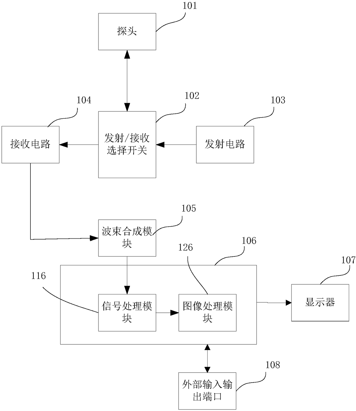
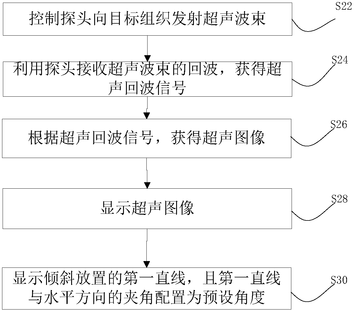

Image

  • Guiding method of ultrasonic detection of pelvic floor and ultrasonic imaging system
  • Guiding method of ultrasonic detection of pelvic floor and ultrasonic imaging system
  • Guiding method of ultrasonic detection of pelvic floor and ultrasonic imaging system

Examples


Embodiment Construction

[0045] The present invention will be further described in detail below through specific embodiments in conjunction with the accompanying drawings. Wherein, similar elements in different implementations adopt associated similar element numbers. In the following implementation manners, many details are described for better understanding of the present application. However, those skilled in the art can readily recognize that some of the features can be omitted in different situations, or can be replaced by other elements, materials, and methods. In some cases, some operations related to the application are not shown or described in the description, this is to avoid the core part of the application being overwhelmed by too many descriptions, and for those skilled in the art, it is necessary to describe these operations in detail Relevant operations are not necessary, and they can fully understand the relevant operations according to the description in the specification and genera...


PUM


Abstract

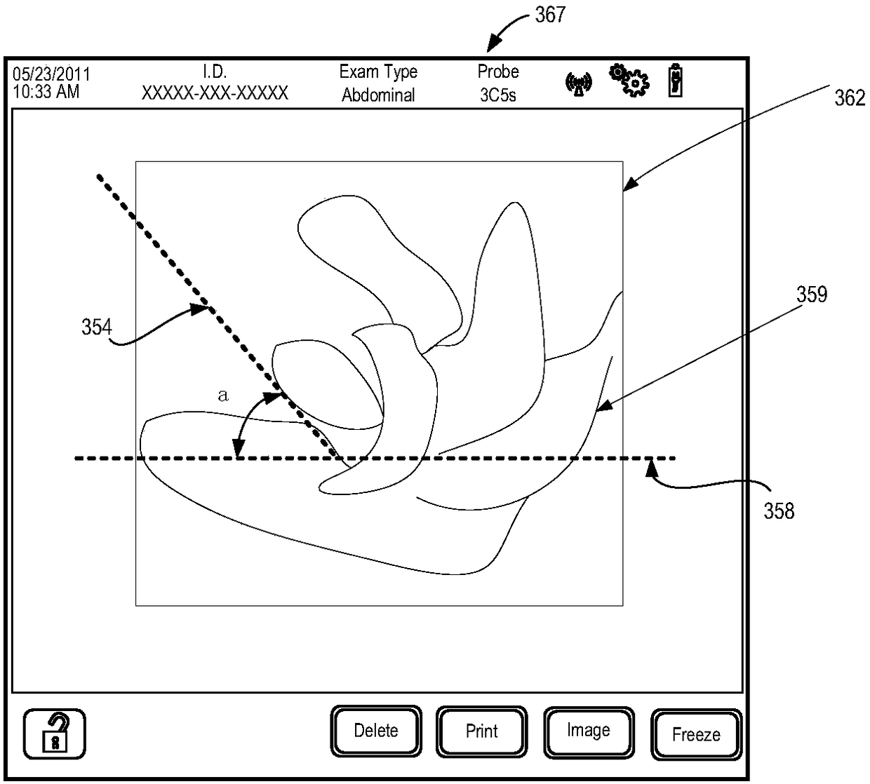
The invention provides a guiding method of ultrasonic detection of a pelvic floor and an ultrasonic imaging system. Therefore, the precision of ultrasound image inspection is increased. The method comprises the following steps: controlling a probe to transmit an ultrasonic beam to a target tissue; utilizing the probe to receive an echo wave of the ultrasonic beams in order to acquire an ultrasonicecho signal; obtaining an ultrasonic image according to the ultrasonic echo signal; and displaying the ultrasonic image; and displaying a first straight line which is disposed in a tilting manner andconfiguring an included angle formed between the first straight line and a horizontal direction to be a pre-set angle, wherein the first straight line is suspended above the ultrasonic image; the pre-set angle is equal to the included angle formed between a horizontal line where the lower margin of a pubic bone is disposed and a combined long axis of the pubic bone.

Description

technical field [0001] The present invention relates to an ultrasonic imaging device, in particular to a guiding method and system provided in the process of image imaging with ultrasonic. Background technique [0002] Female pelvic floor dysfunction is one of the chronic diseases affecting women's daily life, including urinary incontinence, fecal incontinence, recurrent urinary tract infection and pelvic organ prolapse. At present, ultrasonography has gradually replaced MRI as a means of evaluating pelvic floor diseases due to its reproducibility, low cost, simplicity, and no contraindications. In recent years, pelvic floor ultrasound has gradually developed and matured, and has become the preferred imaging method for patients with FPFD. [0003] The ultrasonography industry generally recommends the use of transperineal volume probes or intracavity (transvaginal) volume probes. The transperineal probe is placed on the body surface, so the image display angle is fixed, the...

Claims


Application Information

Patent Timeline
IPC IPC(8): A61B8/08A61B8/12
CPCA61B8/0833A61B8/12A61B8/44A61B8/461A61B8/467A61B8/52
Inventor 温博
Owner SHENZHEN MINDRAY BIO MEDICAL ELECTRONICS CO LTD
Why Eureka
Try Eureka