Preparation method and application of SERS biosensor for detecting tumor marker miRNA-141

A technology of biosensors and tumor markers, applied in the field of detection of tumor marker miRNA-141, can solve the problems of undisclosed SERS biosensors

Active Publication Date: 2018-08-17
NINGBO UNIV
View PDF7 Cites 13 Cited by
  • Summary
  • Abstract
  • Description
  • Claims
  • Application Information

AI Technical Summary

Problems solved by technology

At present, there are no relevant research reports on the preparation method of

Method used


Image

  • Preparation method and application of SERS biosensor for detecting tumor marker miRNA-141
  • Preparation method and application of SERS biosensor for detecting tumor marker miRNA-141
  • Preparation method and application of SERS biosensor for detecting tumor marker miRNA-141

Examples


Embodiment 1

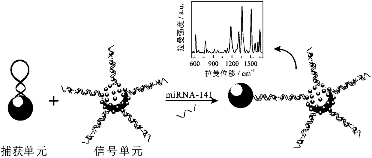
[0040] A method for preparing a SERS biosensor for detecting tumor marker miRNA-141, comprising the following steps:

[0041] (1) Preparation of capture unit

[0042] a. Preparation of Fe 3 o 4 Magnetic nanoparticles solution: 0.8 g FeCl 3 ·6H 2 O and 0.4 g FeCl 2 4H 2 Dissolve O in 70mL of water, pass nitrogen to remove oxygen, stir and mix evenly, raise the temperature to 90 ℃, slowly add 20-25wt% ammonia water dropwise, adjust pH = 9-10, continue heating and stirring for 1-2 h, stop heating, and Under protection, stir and reflux, cool to room temperature, wash with water until neutral, and dilute to 50 mL with ethanol to obtain Fe 3 o 4 magnetic nanoparticle solution;

[0043] b. Preparation of aminated Fe 3 o 4 Magnetic nanoparticles solution: 45 mL Fe 3 o 4 Sonicate the magnetic nanoparticle solution for 0.7 h, add 0.25 mL of 3-aminopropyltriethoxysilane (APTES), stir for 4-5 h, add 1.5 mL of 0.3 mol / L nitric acid solution, continue stirring for 3-4 h, wash wi...

Embodiment 2

[0057] With above-mentioned embodiment 1, its difference is:

[0058] (1) Preparation of capture unit

[0059] a. Preparation of Fe 3 o 4 Magnetic nanoparticles solution: 0.7 g FeCl 3 ·6H 2 O and 0.2 g FeCl 2 4H 2 O dissolved in 50mL of water;

[0060] b. Preparation of aminated Fe 3 o 4 Magnetic nanoparticles solution: 40 mL Fe 3 o 4 Sonicate the magnetic nanoparticle solution for 0.5 h, add 0.2 mL of 3-aminopropyltriethoxysilane, stir for 4 h, add 1 mL of 0.5 mol / L nitric acid solution, and continue stirring for 3 to 4 h;

[0061] c. Preparation of Fe 3 o 4 @Au magnetic nanoparticle solution: 5 mL of aminated Fe 3 o 4 Magnetic nanoparticles solution and 10 mL of 1wt% HAuCl 4 The solution was mixed at an ultrasonic frequency of 60 kHz, and after stirring for 1 h, 30 mL of 30 mmol / L trisodium citrate solution was slowly added and ultrasonicated for 2 h;

[0062] d. Prepare capture unit solution: Take 50 µL Fe 3 o 4 The @Au magnetic nanoparticle solution was s...

Embodiment 3

[0076] With above-mentioned embodiment 1, its difference is:

[0077] (1) Preparation of capture unit

[0078] a. Preparation of Fe 3 o 4Magnetic nanoparticles solution: 1.0 g FeCl 3 ·6H 2 O and 0.5 g FeCl 2 4H 2 O dissolved in 100mL of water;

[0079] b. Preparation of aminated Fe 3 o 4 Magnetic nanoparticles solution: 50 mL Fe 3 o 4 Sonicate the magnetic nanoparticle solution for 1 h, add 0.3 mL of 3-aminopropyltriethoxysilane, stir for 5 h, add 2 mL of 0.1 mol / L nitric acid solution, and continue stirring for 3 to 4 h;

[0080] c. Preparation of Fe 3 o 4 @Au magnetic nanoparticle solution: 10 mL of aminated Fe 3 o 4 Magnetic nanoparticles solution and 20 mL of 1wt% HAuCl 4 The solution was mixed at an ultrasonic frequency of 100 kHz, and after stirring for 1 h, 50 mL of 20 mmol / L trisodium citrate solution was slowly added, and ultrasonicated for 2 to 3 h;

[0081] d. Prepare capture unit solution: Take 100 µL Fe 3 o 4 The @Au magnetic nanoparticle solutio...


PUM


Abstract

The invention discloses a preparation method and an application of a SERS biosensor for detecting a tumor marker miRNA 141. The method comprises the following steps: (1) combining a Fe3O4@Au magneticnanoparticle with a capture probe to obtain a capture unit solution; (2) combining a hollow gold nanosphere with an initiation probe to prepare a signal unit solution; and (3) reacting and combining acapture unit, miRNA 141 and a signal unit, and performing magnetic separation to obtain the SERS biosensor. The application is characterized in that an SERS signal is detected by a Raman spectrometer, SERS signal intensities corresponding to a series of miRNA-141 with different concentrations are detected, and a quantitative relationship between the concentration of the miRNA-141 and the SERS signal intensity is established; and the concentration of miRNA-141 in an unknown sample is detected according to the quantitative relationship. The SERS biosensor has the advantages of high sensitivity,high specificity, simple assembling process and fast detection speed.

Description

technical field [0001] The invention relates to a detection method for detecting tumor marker miRNA-141, in particular to a preparation method and application of a SERS biosensor for detecting tumor marker miRNA-141. Background technique [0002] Cancer, also known as malignant tumor, is essentially a polygenic disease caused by abnormal cell growth and proliferation mechanism, which seriously endangers human health. The International Agency for Research on Cancer under the United Nations World Health Organization predicts that 13.2 million people will die of cancer by 2030, and the number of confirmed cases will reach 21.4 million. Although the mortality rate of cancer is high, it is not incurable. Studies have shown that early cancer is the best time to cure cancer. Therefore, early detection, early diagnosis and early treatment of tumors can effectively prevent and treat cancer. The discovery of tumor markers makes early diagnosis possible. Tumor markers are substances ...

Claims


Application Information

Patent Timeline
IPC IPC(8): G01N33/574
CPCG01N33/57484
Inventor 卢静郭智勇贾亚茹邵慧丽胡宇芳王邃
Owner NINGBO UNIV
Why Eureka
Try Eureka