CT image pulmonary nodule detection method, device, equipment and readable storage medium

A CT image and detection method technology, applied in the field of image processing, can solve problems such as poor accuracy of pulmonary nodules

Active Publication Date: 2021-02-09
SHENZHEN IMSIGHT MEDICAL TECH CO LTD
View PDF5 Cites 0 Cited by
  • Summary
  • Abstract
  • Description
  • Claims
  • Application Information

AI Technical Summary

Problems solved by technology

[0003] The main purpose of the present invention is to provide a method, device, equipment and readable storage medium for detecting pulmonary nodules in CT images, aiming to solve the technical problem in the prior art that the accuracy of automatic detection of pulmonary nodules based on CT images is not good

Method used


Image

  • CT image pulmonary nodule detection method, device, equipment and readable storage medium
  • CT image pulmonary nodule detection method, device, equipment and readable storage medium
  • CT image pulmonary nodule detection method, device, equipment and readable storage medium

Examples


Embodiment Construction

[0081] It should be understood that the specific embodiments described here are only used to explain the present invention, not to limit the present invention.

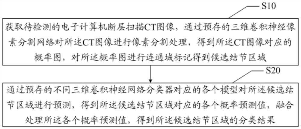
[0082] The present invention provides a method for detecting pulmonary nodules in CT images. In the first embodiment of the method for detecting pulmonary nodules in CT images of the present invention, refer to figure 1 , the CT image pulmonary nodule detection method comprises:

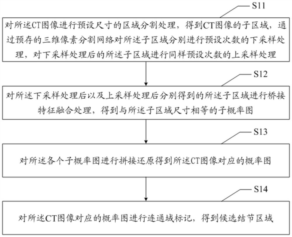
[0083] Obtain a computer tomography CT image to be detected, perform pixel segmentation processing on the CT image through a pre-stored three-dimensional convolutional neural pixel segmentation network, obtain a probability map corresponding to the CT image, and perform connected domain marking on the probability map Obtain the candidate nodule area; predict the candidate nodule area through each prediction model corresponding to the different pre-stored three-dimensional convolutional neural network classifiers, obtain each probability pred...


PUM


Abstract

The invention discloses a method, device, equipment and readable storage medium for detecting pulmonary nodules in CT images. The method for detecting pulmonary nodules in CT images comprises: acquiring a computerized tomography CT image to be detected, and using a pre-stored three-dimensional volume The neural pixel segmentation network performs pixel segmentation processing on the CT image to obtain a probability map corresponding to the CT image, and performs connected domain marking on the probability map to obtain candidate nodule regions; classify through different pre-stored three-dimensional convolutional neural networks A plurality of models corresponding to the device predict the candidate nodule region to obtain respective probability prediction values ​​corresponding to the candidate nodule region, and fuse and process the respective probability prediction values ​​to obtain a classification result of the candidate nodule region. The invention solves the existing technical problem of poor accuracy of automatic detection of pulmonary nodules based on CT images.

Description

technical field [0001] The present invention relates to the technical field of image processing, in particular to a method, device, equipment and readable storage medium for detecting pulmonary nodules in CT images. Background technique [0002] Pulmonary nodules are one of the most important early signs of lung cancer. According to the lesion characteristics of lung nodules, the lesion characteristics of lung lesions can be inferred. Due to the uncertainty of the size, shape and density of nodules, existing detection methods are difficult to detect. To meet the market demand for the accuracy of pulmonary nodule detection. Contents of the invention [0003] The main purpose of the present invention is to provide a method, device, equipment and readable storage medium for detecting pulmonary nodules in CT images, aiming to solve the technical problem in the prior art that the accuracy of automatic detection of pulmonary nodules based on CT images is not good. [0004] In o...

Claims


Application Information

Patent Timeline
Patent Type & Authority Patents(China)
IPC IPC(8): G06T7/00G06T7/11G06T7/187G06T3/40G06V10/26G06V10/50G06V10/764G06V10/774
CPCG06T3/4038G06T7/0012G06T7/11G06T7/187G06T2207/20081G06T2207/20084G06T2207/10081G06T2207/20221G06T2207/30064A61B6/5217A61B6/032G06T7/143G16H50/30G06V10/26G06V10/50G06V10/454G06V2201/03G06V10/82G06V10/764G06V10/774G06F18/2413A61B6/50A61B6/5205G06T7/0016G06T2207/20076G06F18/217G06F18/2415
Inventor 窦琪刘权德陈浩
Owner SHENZHEN IMSIGHT MEDICAL TECH CO LTD
Why Eureka
Try Eureka