A Segmentation Method of CT Chest X-ray Lung Tissue Image Based on Level Set
An image segmentation and CT image technology, applied in the field of medical image processing, can solve the problems of easy omission of lung nodules, inaccurate segmentation boundaries, and dependence on segmentation templates, and achieve the effects of accurate segmentation results, high precision, and strong robustness.
- Summary
- Abstract
- Description
- Claims
- Application Information
AI Technical Summary
Problems solved by technology
Method used
Image
Examples
Embodiment 1
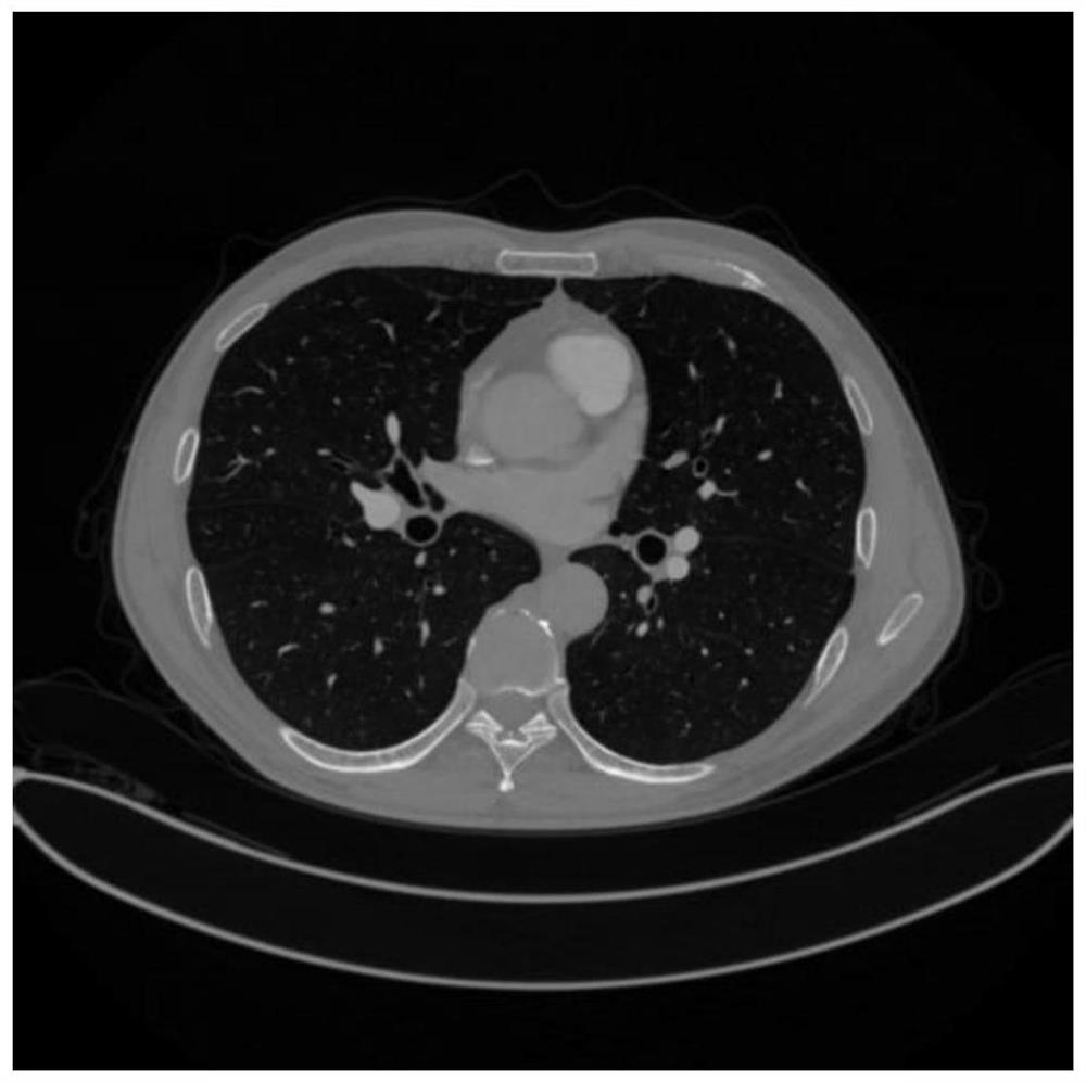
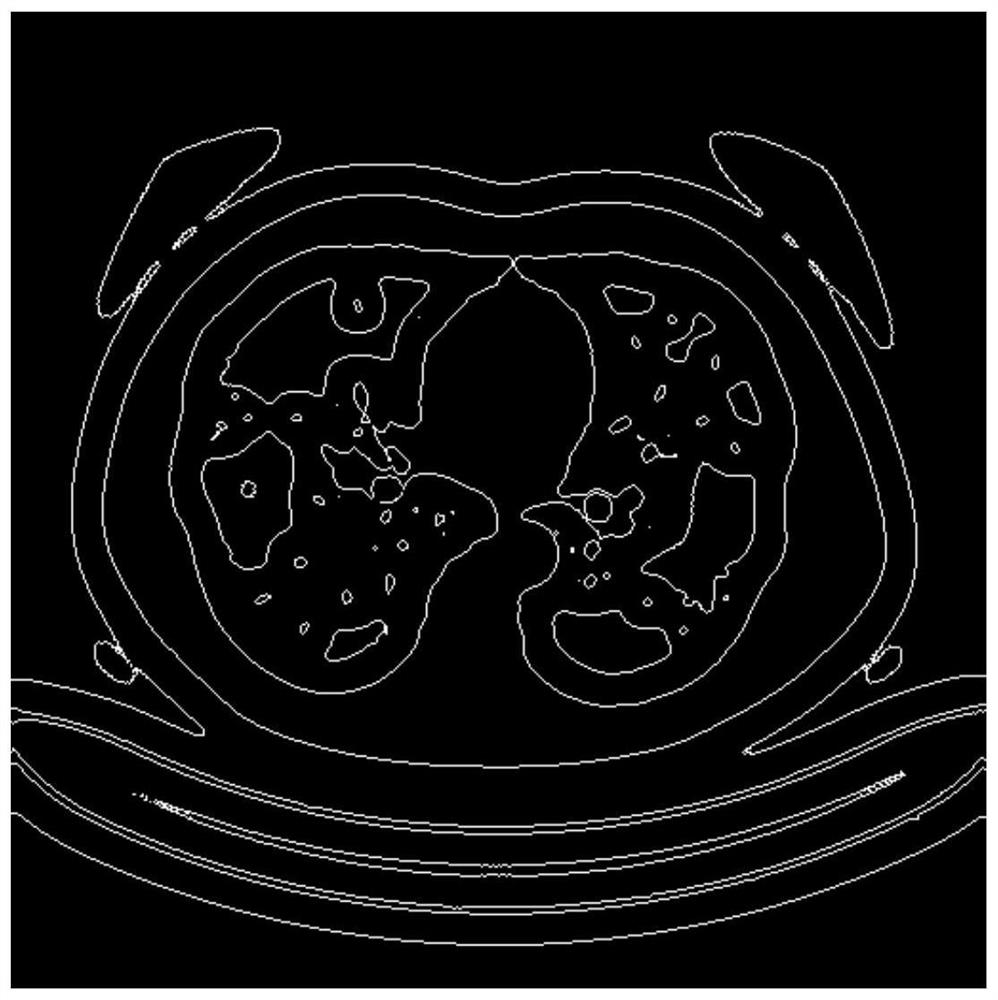
[0027] The results of the existing CT chest X-ray lung tissue image segmentation methods are not robust enough, rely heavily on the segmentation template, the segmentation boundary is not precise enough, and the segmentation process requires manual intervention. Aiming at the shortcomings of existing methods, a robust, fully automatic and accurate method for lung tissue image segmentation on CT chest X-rays is needed. For this reason the present invention proposes a kind of CT chest X-ray lung tissue image segmentation method based on level set, see figure 2 , including the following steps:
[0028] Step 1 Obtain a chest CT image containing lung tissue, use MATLAB to read the image into the computer, store the image in the computer as a three-dimensional tensor, and perform CT chest image preprocessing. The method of image preprocessing is to give the CT image grayscale Add 400 to the value, so that the distribution of the gray histogram of the original input CT image can be...
Embodiment 2
[0036] Level set-based CT chest X-ray lung tissue image segmentation method is the same as embodiment 1-1, see figure 2 , because the grayscale value dynamic range of the original input CT image is -1024 to +1024, and the image grayscale range of the statistical lung area is -600 to -200, so the image preprocessing described in step 1 is for CT Add 400 to the grayscale value of the image, so that the grayscale range of the image in the lung area can be adjusted to -200 to +200, making the lung area more prominent and convenient for subsequent processing. In order to reduce the complexity of the algorithm and reduce the interference of the image area other than the lung area, the gray value of the image is normalized to an 8-bit unsigned number.
Embodiment 3
[0038] The CT chest X-ray lung tissue image segmentation method based on the level set is the same as embodiment 1-2, and the energy functional equation is constructed on the preprocessed CT chest X-ray lung tissue image described in step 2 and an initial zero level set is set. , including the following steps:
[0039] 2.1 Construct the energy functional equation F(φ,c,b)
[0040] F(φ,c,b)=ε(φ,c,b)+υL(φ)+μR p (φ)
[0041] Where ε(φ,c,b) is the energy function, φ is the level set function, and c is the gray average vector inside and outside the level set, because CT images have different radioactivity levels, different imaging equipment, and different environment noise, so the bias field correction is introduced when constructing the energy functional equation. The bias field correction can reduce the influence of the above factors on image segmentation. b is the bias field parameter. At the same time, when constructing the energy functional equation The zero level set arc l...
PUM
 Abstract
Description
Claims
Application Information
 - R&D
- Intellectual Property
- Life Sciences
- Materials
- Tech Scout
- Unparalleled Data Quality
- Higher Quality Content
- 60% Fewer Hallucinations
Browse by: Latest US Patents, China's latest patents, Technical Efficacy Thesaurus, Application Domain, Technology Topic, Popular Technical Reports.
© 2025 PatSnap. All rights reserved.Legal|Privacy policy|Modern Slavery Act Transparency Statement|Sitemap|About US| Contact US: help@patsnap.com


