A pathological image quality validity identification method

A technology of pathological images and identification methods, applied in image enhancement, image analysis, image data processing, etc., can solve the problems of uneven image quality of pathological slices, uneven levels of hospital operators, and inability to identify slice pathologists. , to achieve the effect of fully automated processing, objective and stable judgment, and reduced workload

Active Publication Date: 2019-01-11
GENERAL HOSPITAL OF PLA
View PDF7 Cites 18 Cited by
  • Summary
  • Abstract
  • Description
  • Claims
  • Application Information

AI Technical Summary

Problems solved by technology

Although the country has very strict standards for pathological slices, in the implementation process of specific hospitals, due to the uneven level of hospital operators and various errors in the process of implementation, the image quality of the final imaging pathological slices is un

Method used


Image

  • A pathological image quality validity identification method
  • A pathological image quality validity identification method
  • A pathological image quality validity identification method

Examples


Example Embodiment

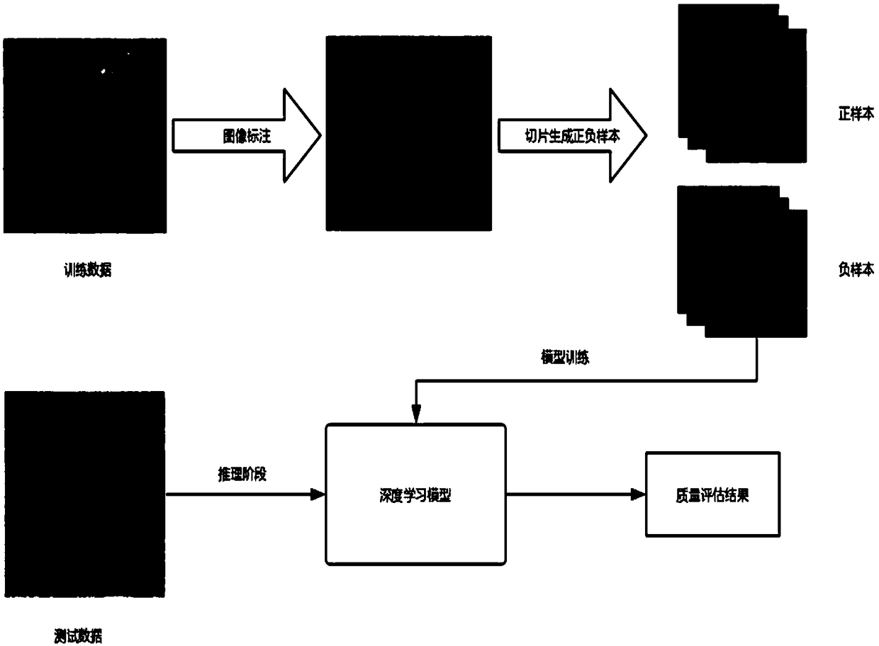
[0023] The specific process of the present invention will be described below by taking skin pathological slices as an example.

[0024] Because some areas of the image may be very blurred due to contamination, over-staining or tissue folding in the slice imaging, the cell morphology cannot be seen clearly for subsequent diagnosis.

[0025] like figure 1 Shown, the present invention is concretely realized as follows:

[0026] (1) First use binarization filtering to remove the white background;

[0027] (2) In the low-magnification mirror, the doctor marks the areas that meet the imaging quality requirements and the areas that do not meet the imaging quality;

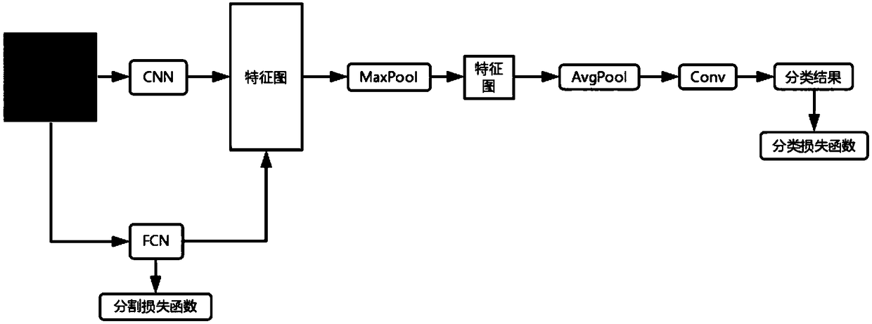
[0028] (3) After collecting enough labeled data and qualified and non-compliant regions marked by doctors, use convolutional network to construct binary classification network to classify the quality of imaging. Ignore the white background during model training and only focus on training the network in the tissue area;...


PUM


Abstract

The invention relates to a pathological image quality validity identification method, which collects pathological images and labels the quality of imaging pathological slices in the pathological images to form labeling information. According to the labeling information, the regions with good image quality and the regions with bad image quality are obtained. The regions with good image quality refer to the regions where the cell outline shape is very clear, and such regions are the regions with good image quality. The poor image quality region refers to some regions which are very blurry, and these regions are the poor image quality regions; the two types of sample data are trained to obtain a trained binary classifier; the binary classifier is used to analyze the data of pathological images and get the analysis result chart; according to the judgment rules, whether the pathological slices in the pathological images meet the requirements of clinical doctors are determined. The inventioncan greatly reduce the time cost in the process of sending and reading slices back and forth due to the slice making problem, and improve the efficiency from the slice making to the diagnosis.

Description

technical field [0001] The invention relates to a method for identifying the validity of pathological image quality, which belongs to the technical field of artificial intelligence. Background technique [0002] Pathological slide analysis is a method for judging diseases based on cell shape and pattern, which is made by cutting tissue with precision instruments and then going through multiple processes. Although the country has very strict standards for pathological slices, in the implementation process of specific hospitals, due to the uneven level of hospital operators and various errors in the process of implementation, the image quality of the final imaging pathological slices is uneven, and even some Slices cannot be recognized by pathologists due to process problems, which is seriously the work of imaging pathologists. [0003] At present, there is no suitable fully automated tool and method for judging the imaging quality of pathological image slices. Contents of ...

Claims


Application Information

Patent Timeline
IPC IPC(8): G06T7/00
CPCG06T7/0012G06T2207/20221G06T2207/30168G06T2207/20081G06T2207/20084
Inventor 巴伟李承新王彬华邹进屹
Owner GENERAL HOSPITAL OF PLA
Why Eureka
Try Eureka