A method for detecting a beam limiter region in a breast image, a method for determining a boundary in that breast image, and a medical device

A beam limiter and image-in-the-image technology, applied in the field of medical image processing, can solve the problems of reducing breast image post-processing effect, increasing breast image post-processing complexity and difficulty, mechanical position error, etc.

Active Publication Date: 2019-01-18
SHANGHAI UNITED IMAGING HEALTHCARE
View PDF11 Cites 8 Cited by
  • Summary
  • Abstract
  • Description
  • Claims
  • Application Information

AI Technical Summary

Problems solved by technology

[0003] However, due to defects such as mechanical position errors, the beam limiter area will still remain in the cropped breast image, and since the beam limiter is a high-attenuation material, the re

Method used


Image

  • A method for detecting a beam limiter region in a breast image, a method for determining a boundary in that breast image, and a medical device
  • A method for detecting a beam limiter region in a breast image, a method for determining a boundary in that breast image, and a medical device
  • A method for detecting a beam limiter region in a breast image, a method for determining a boundary in that breast image, and a medical device

Examples


Embodiment Construction

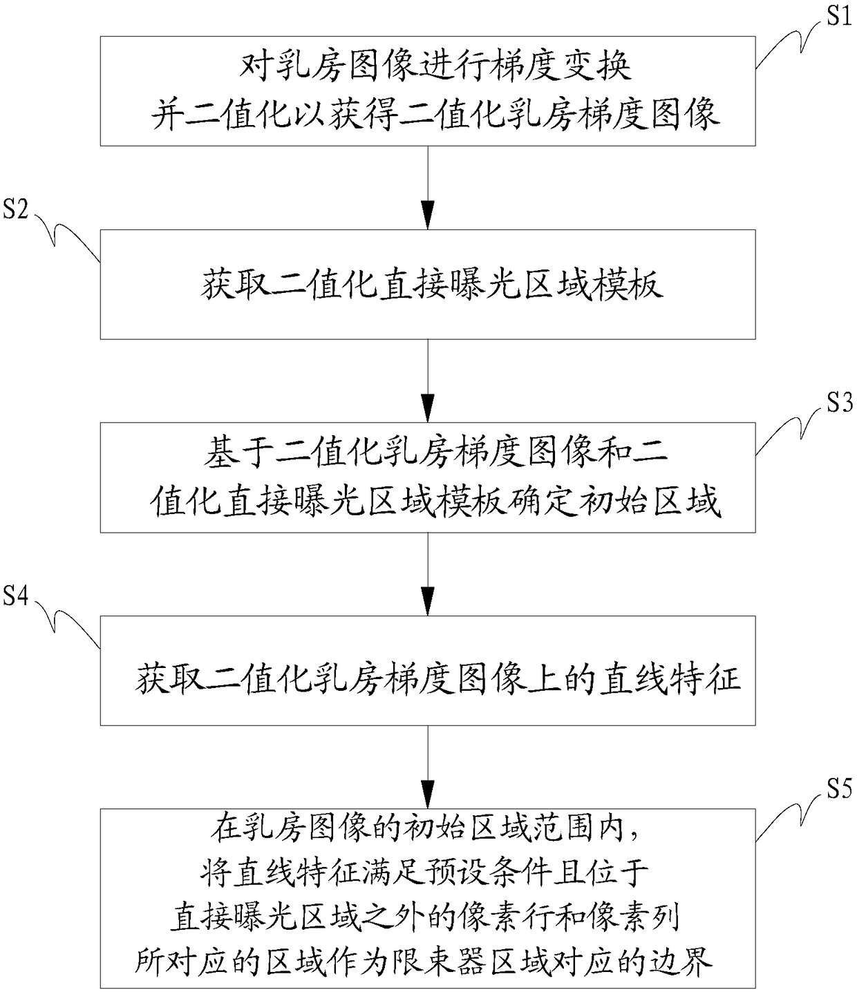
[0180] In order to make the object, technical solution and advantages of the present invention clearer, the present invention will be further described in detail below in conjunction with the accompanying drawings and embodiments. It should be understood that the specific embodiments described here are only used to explain the present invention, not to limit the present invention.

[0181] During the image capture operation on the compressed breast tissue, there may be a beam limiter area in the captured breast image, and since the above-mentioned beam limiter is a high-attenuation material, the corresponding range in the above-mentioned breast image will be There is a high attenuation area inside, and the high attenuation area will increase the complexity and difficulty of the post-processing of the breast image, thereby reducing the post-processing effect of the breast image. Traditionally, the high attenuation area corresponding to the beam limiter in the breast image is re...


PUM


Abstract

The invention relates to the technical field of medical image processing, in particular to a method for detecting a beam limiter region in a breast image, a method for determining a boundary in the breast image and a medical device. According to the method for detecting a beam limiter region in a breast image, Through gradient transformation and binarization of breast images, and further based onthe binary breast gradient image obtained after binarization and the binary direct exposure region template, an initial region in the breast image is determined, by combining the linear features of binary breast gradient images, the accurate positioning of the boundary of the beam limiter region in the breast image can be realized, thereby ensuring that the breast image clipped based on the positioned boundary does not remain the finite beam region, so as to effectively reduce the complexity and difficulty of the breast image post-processing and improve the post-processing effect of the breastimage.

Description

technical field [0001] The invention relates to the technical field of medical image processing, in particular to a method for detecting a beam limiter area in a breast image, a method for determining boundaries in the breast image and medical equipment. Background technique [0002] When using the Digital Mammography System (Digital Mammography System, DMG for short) to scan and shoot breast images, according to clinical needs, the corresponding imaging tissue can be compressed by selecting different compression plates, and the size of the opening of the breast beam limiter can be adjusted. , so that the size of the opening of the breast beam limiter is consistent with the size of the compression plate used, and then the captured breast image can be cropped according to the coordinates of the mechanical feedback to remove the beam limiter area in the breast image. [0003] However, due to defects such as mechanical position errors, the beam limiter area will still remain in...

Claims


Application Information

Patent Timeline
IPC IPC(8): G06T7/00G06T7/12G06T5/20G06T5/30G06T3/40G06K9/38
CPCG06T3/4007G06T5/20G06T5/30G06T7/0012G06T7/12G06T2207/10116G06T2207/20032G06T2207/30068G06T2207/20132G06V10/28
Inventor 闫晶王汉禹牛杰
Owner SHANGHAI UNITED IMAGING HEALTHCARE
Why Eureka
Try Eureka