
Oral diagnostic device and image processing method

A diagnostic device and image processing technology, applied in the field of dental instruments, can solve the problems of inability to diagnose immediately at home, inability to self-diagnose patients, and inability to carry it around, so as to improve ease of use and work efficiency, efficient judgment, and simplify complex processes Effect

Inactive Publication Date: 2019-01-29
CHANGAN UNIV
View PDF5 Cites 1 Cited by
  • Summary
  • Abstract
  • Description
  • Claims
  • Application Information

AI Technical Summary

Problems solved by technology

At present, oral treatment and diagnosis must go to the hospital for oral image collection before treatment can be performed, and immediate diagnosis cannot be performed at home
The traditional oral image acquisition equipment is completed by the oral mirror, and it needs to be transmitted to the computer for viewing through the data cable. It is large in size and can only be used in the hospital. It cannot be carried around and cannot allow patients to self-diagnose

Method used


Image

  • Oral diagnostic device and image processing method
  • Oral diagnostic device and image processing method
  • Oral diagnostic device and image processing method

Examples


Embodiment Construction

[0043] The present invention will be further described below in conjunction with accompanying drawing.

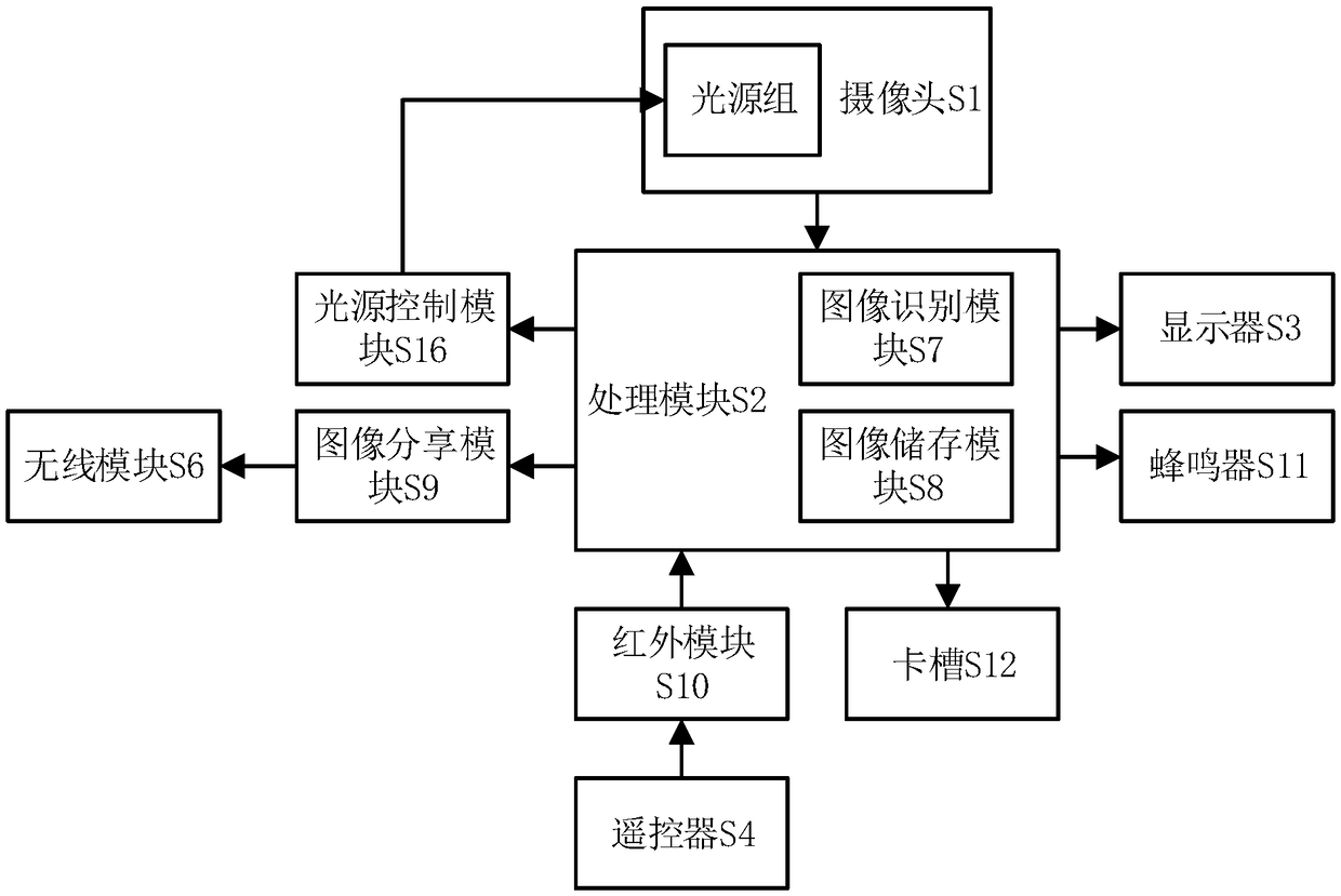
[0044] see figure 1 , a kind of oral cavity diagnosis device, it is characterized in that, comprises camera S1, and camera S1 is connected processing module S2, and processing module S2 comprises image recognition module S7 and image storage module S8, and processing module S2 is connected display S3, and camera S1 is provided with light source group , the light source group is controlled by the light source control module S16, the light source control module S16 is connected to the processing unit S2, the light source controller S16 is used to collect intraoral images in the processing unit S2, and controls the brightness of the light source group according to the brightness of the image.

[0045] The camera S1 is used to collect intraoral images, and send the collected intraoral images to the processing module S2;

[0046] The image recognition module S7 is used for segm...


PUM


Abstract

The invention discloses an oral diagnostic device and an image processing method. The oral diagnostic device comprises a camera; the camera is connected to a processing module; the processing module comprises an image recognition module and an image storage module, and the processing module is connected to a display. The camera is used to acquire images, and the image device module can process theimages to mark images with abnormal conditions, the complicated process of traditional oral inspection is simplified, and the usability and the working efficiency can be improved. The device enablesthe patient to diagnose the oral problem at home, and is easy to carry, so that the patient can seek medical treatment in time.

Description

technical field [0001] The invention relates to the field of oral instruments, in particular to an oral diagnosis device and an image processing method. Background technique [0002] According to the changes in life in recent years, oral problems have been paid more and more attention. At present, oral treatment and diagnosis must go to the hospital for oral image collection before treatment can be performed, and immediate diagnosis cannot be performed at home. The traditional oral image acquisition equipment is completed by oral speculum, and it needs to be transmitted to the computer for viewing through the data cable. It is large in size and can only be used in hospitals, and cannot be carried around, and cannot allow patients to self-diagnose. Contents of the invention [0003] The purpose of the present invention is to overcome the above-mentioned shortcomings, and provide an oral diagnosis device and image processing method, so that patients can self-diagnose oral p...

Claims


Application Information

Patent Timeline
Patent Type & Authority Applications(China)
IPC IPC(8): G16H50/20G06K9/62G06N3/08G06T7/136
CPCG06N3/08G06T7/136G16H50/20G06T2207/30036G06F18/232G06F18/2135G06F18/2411G06F18/214
Inventor 王春梅王江安贺永杰王凯李鑫
Owner CHANGAN UNIV
Learn More