Hepatic space-occupying lesion identification method and device and implement device

An identification method and technology for venereal diseases, applied in the field of medical imaging, can solve the problems of inability to consider the horizontal features of four-stage images and the longitudinal features of single-stage images, and the low accuracy of identification results.

Active Publication Date: 2019-03-08
BEIJING PEREDOC TECH CO LTD
View PDF9 Cites 9 Cited by
  • Summary
  • Abstract
  • Description
  • Claims
  • Application Information

AI Technical Summary

Problems solved by technology

[0003] At present, liver space-occupying lesions are mainly identified manually from CT images, and automatic computer identification is mostly based on single-phase images of CT images, which cannot consider the horizontal characteristics between the four-phase images and the longitudinal characteristics of a single-phase image. The accuracy of the recognition result is not high

Method used


Image

  • Hepatic space-occupying lesion identification method and device and implement device
  • Hepatic space-occupying lesion identification method and device and implement device
  • Hepatic space-occupying lesion identification method and device and implement device

Examples


Embodiment Construction

[0044]In order to make the purpose, technical solutions and advantages of the embodiments of the present invention clearer, the technical solutions of the present invention will be clearly and completely described below in conjunction with the accompanying drawings. Obviously, the described embodiments are part of the embodiments of the present invention, not all of them. the embodiment. Based on the embodiments of the present invention, all other embodiments obtained by persons of ordinary skill in the art without making creative efforts belong to the protection scope of the present invention.

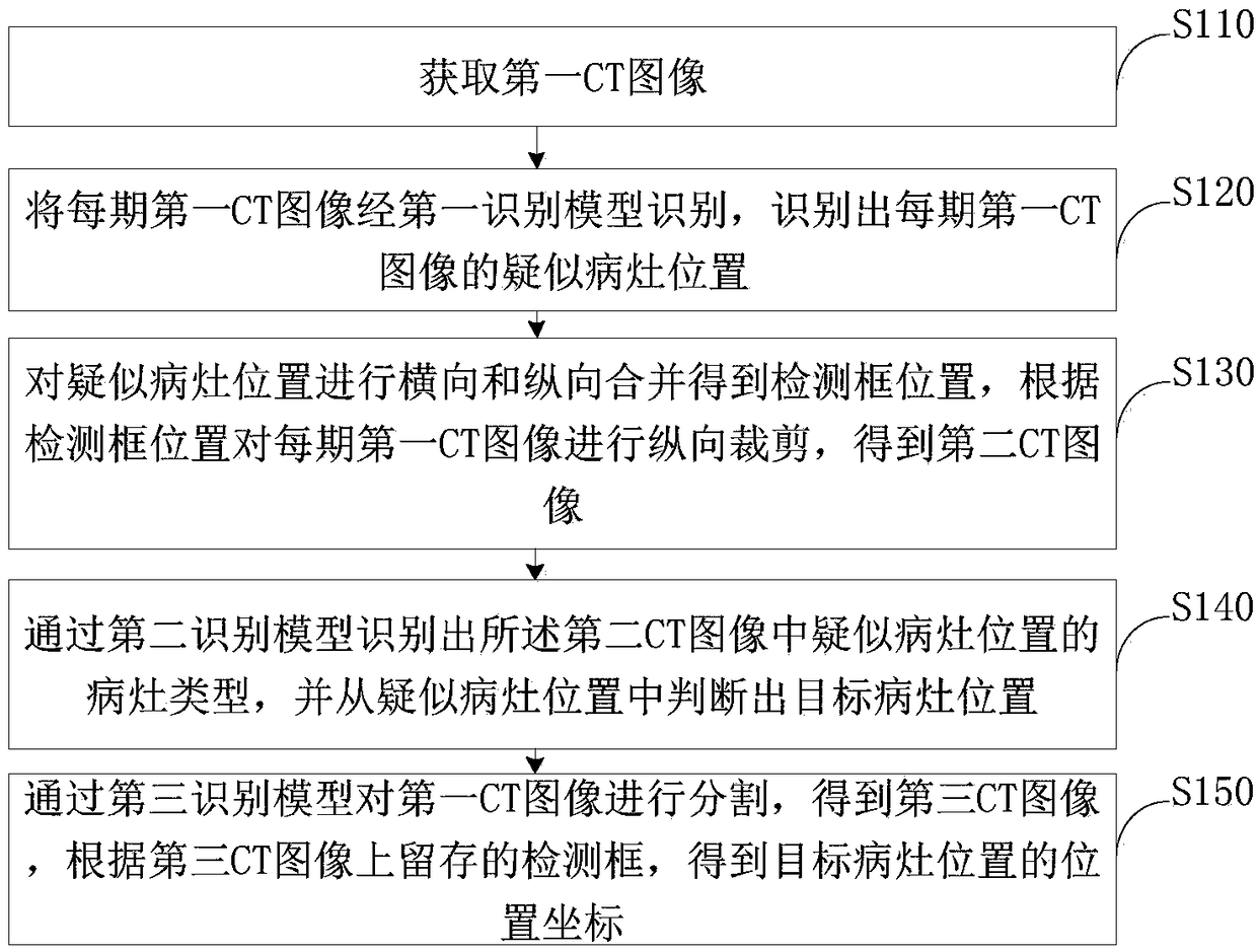
[0045] At present, liver space-occupying lesions are mainly identified manually from CT images, and automatic computer identification is mostly based on single-phase images of CT images, which cannot consider the horizontal features between the four-phase images and the longitudinal features of a single-phase image. The accuracy of the recognition result is not high.

[0046] Based o...


PUM


Abstract

The invention provides a liver space-occupying lesion identification method and device and an implementation device, which relate to the technical field of medical images. The method comprises the steps of acquiring a first CT image; recognizing the first CT image of each phase by a first recognition model, and recognizing the suspected focus position of the first CT image of each phase; carryingout the lateral and longitudinal combination of suspect lesion positions to obtain a detection frame position, and cutting that first CT image of each phase longitudinally according to the detection frame position to obtain a second CT image; identifying the lesion type of the suspected lesion position in the second CT image through the second recognition model, and judging the target lesion position from the suspected lesion position; segmenting the first CT image by the third recognition model, and obtaining the third CT image; and according to the detection frame on the third CT image, obtaining the position coordinates of the target focus. The lateral features between the four-phase images and the longitudinal features of the single-phase images are comprehensively considered, which greatly improves the accuracy of the recognition results.

Description

technical field [0001] The invention relates to the technical field of medical imaging, in particular to a method, device and implementation device for identifying liver space-occupying lesions. Background technique [0002] Space-occupying lesion is a special term in medical imaging diagnostics. It usually appears in X-ray, B-ultrasound, CT and other examination results. , arterial phase, portal venous phase and delayed phase four-phase images. [0003] At present, liver space-occupying lesions are mainly identified manually from CT images, and automatic computer identification is mostly based on single-phase images of CT images, which cannot consider the horizontal characteristics between the four-phase images and the longitudinal characteristics of a single-phase image. The accuracy of the recognition result is not high. Contents of the invention [0004] In view of this, the purpose of the present invention is to provide a liver space-occupying lesion identification ...

Claims


Application Information

Patent Timeline
Patent Type & Authority Applications(China)
IPC IPC(8): G06T7/00G06T7/11G06T7/187
CPCG06T7/0012G06T2207/10081G06T2207/20081G06T2207/20084G06T2207/30056G06T7/11G06T7/187
Inventor 许志方王方
Owner BEIJING PEREDOC TECH CO LTD
Why Eureka
Try Eureka