
A medical image segmentation method

A medical image and image technology, applied in the field of computer vision technology and medical image analysis, can solve problems such as mutual adhesion of similar objects, and achieve the effect of ensuring robustness and reliability, solving mutual adhesion, and the method is simple and efficient

Inactive Publication Date: 2019-03-29
SHENZHEN GRADUATE SCHOOL TSINGHUA UNIV
View PDF8 Cites 9 Cited by
  • Summary
  • Abstract
  • Description
  • Claims
  • Application Information

AI Technical Summary

Problems solved by technology

[0006] In view of the above technical problems, the present invention proposes a medical image segmentation method, which uses instance segmentation to achieve accurate segmentation of each region of interest in the medical image, and solves the problem of similar objects sticking to each other in the existing segmentation methods. The method is simple and efficient

Method used


Image

  • A medical image segmentation method
  • A medical image segmentation method
  • A medical image segmentation method

Examples


Embodiment Construction

[0033] The present invention will be further described below with reference to the accompanying drawings and in combination with preferred embodiments.

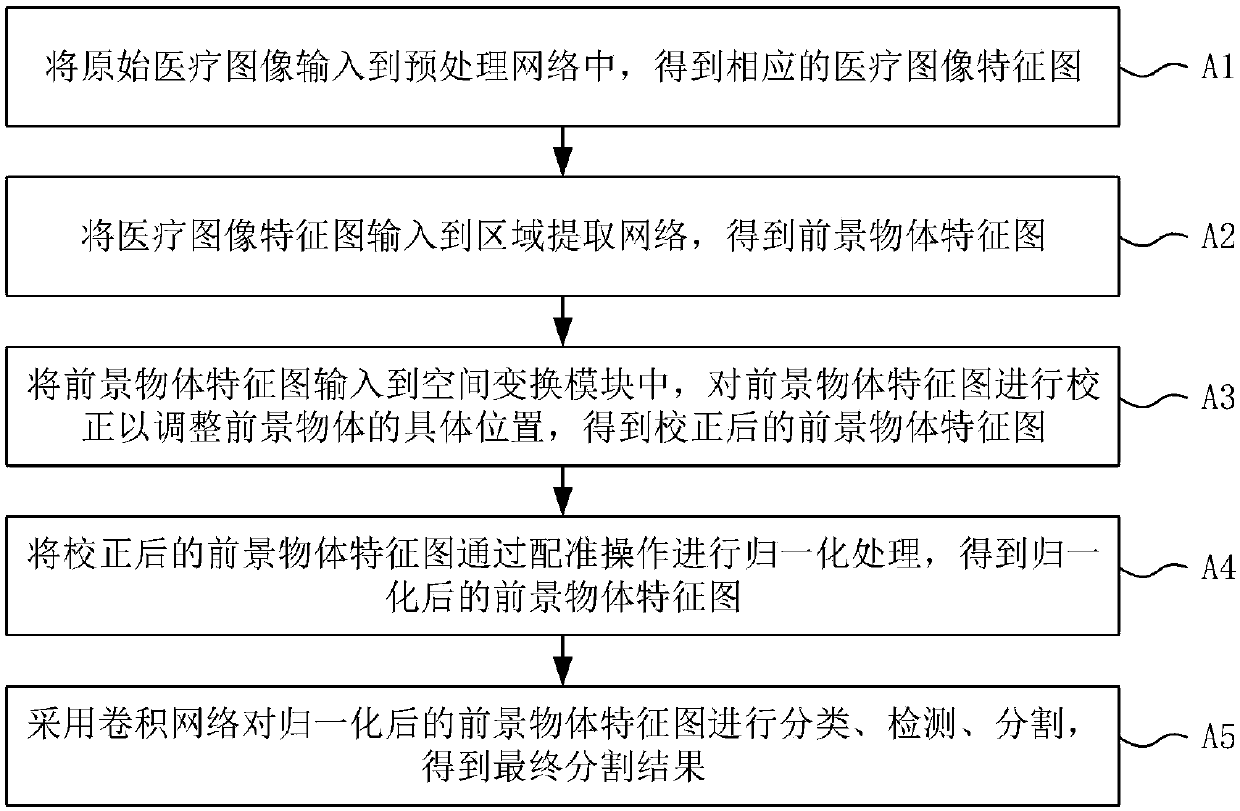
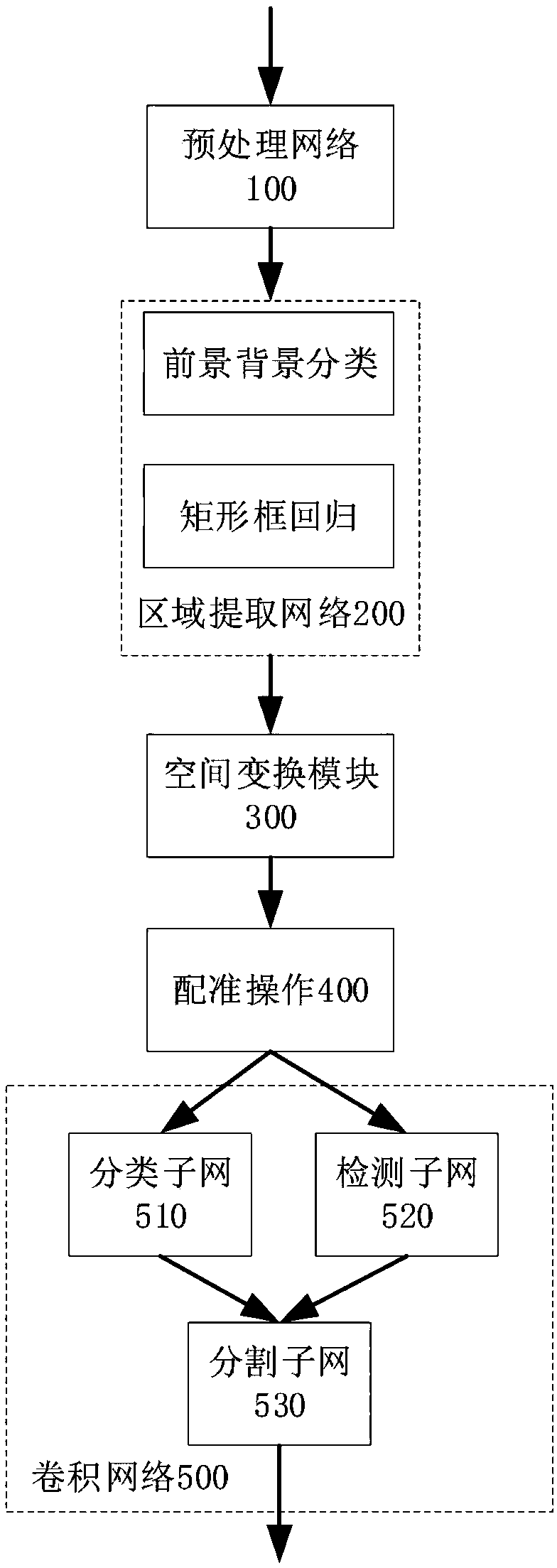
[0034] combine figure 1 and figure 2 , the preferred embodiment of the present invention discloses an accurate and robust medical image segmentation method, comprising the following steps:

[0035] A1: Input the original medical image into the preprocessing network 100 to obtain the corresponding feature map;

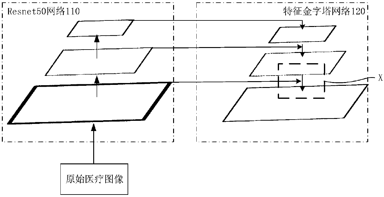
[0036] like image 3 , the preprocessing network includes a Resnet50 network 110 and a feature pyramid network 120. First, the original medical image is input to the Resnet50 network 110 on the left side of the figure. After processing, the processing result of the Resnet50 network 110 is input to the feature pyramid network 120 on the right side of the figure. middle.

[0037] Among them, the Resnet50 network has the advantages of image feature extraction and the characteristics of simple and easy training of the...


PUM


Abstract

The invention discloses a medical image segmentation method, comprising the following steps: A1, inputting an original medical image into a preprocessing network to obtain a corresponding medical image characteristic map; A2, inputting that medical image feature map to an area extraction network to obtain all foreground object feature maps; A5: adopting a convolution network to classify, detect and segment that feature map of the foreground object to obtain the final segmentation result. The medical image segmentation method provided by the invention realizes the accurate segmentation of eachregion of interest of the medical image by adopting an example segmentation, solves the problem that similar objects adhere to each other in the prior segmentation method, and has the advantages of simple and high efficiency.

Description

technical field [0001] The invention relates to the fields of computer vision technology and medical image analysis, in particular to a medical image segmentation method. Background technique [0002] Deep learning has achieved great success in the field of computer vision in recent years, which mainly includes basic tasks such as image recognition, target detection and segmentation, as well as high-level tasks such as gesture recognition, pedestrian re-identification, and question-answering systems. In many tasks, the performance of deep learning has surpassed that of humans, which greatly liberated manpower. In the medical field, the development of the field of medical image analysis has been relatively slow due to the particularity of the disease, the long training period for doctors, and the low error tolerance rate. Therefore, the performance of computer-aided diagnosis technology based on medical image analysis also needs to be improved. [0003] Domestic medical reso...

Claims


Application Information

Patent Timeline
IPC IPC(8): G06T7/11
CPCG06T2207/10004G06T2207/20081G06T2207/20084G06T2207/30004G06T7/11
Inventor 王好谦宋磊张永兵戴琼海
Owner SHENZHEN GRADUATE SCHOOL TSINGHUA UNIV
Learn More