Implant and kit for treatment of bone lesion site, as well as method for treating bone lesion site

A technology for implants and bone injuries, applied in the direction of bone implants, chemical instruments and methods, drug combinations, etc., can solve the problems of unreported bone injury healing, damage and defect or complex shape, etc., to promote bone formation , improve the proliferation ability, and promote the effect of treatment

Inactive Publication Date: 2019-04-02
UBE IND LTD +1
View PDF9 Cites 3 Cited by
  • Summary
  • Abstract
  • Description
  • Claims
  • Application Information

AI Technical Summary

Problems solved by technology

In these methods, there are cases where it is not suitable for dealing with large damages and defects or complex shapes, and it is very troublesome.
In addition, it has not been reported that the above-mentioned biomaterials or bioabsorbable materials themselves promote the healing of bone injuries

Method used


Image

  • Implant and kit for treatment of bone lesion site, as well as method for treating bone lesion site
  • Implant and kit for treatment of bone lesion site, as well as method for treating bone lesion site
  • Implant and kit for treatment of bone lesion site, as well as method for treating bone lesion site

Examples


Embodiment 1

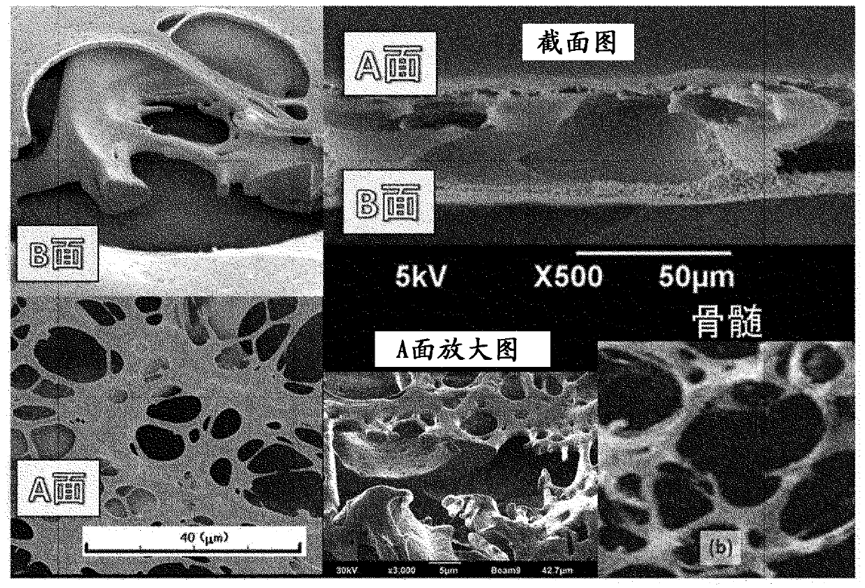
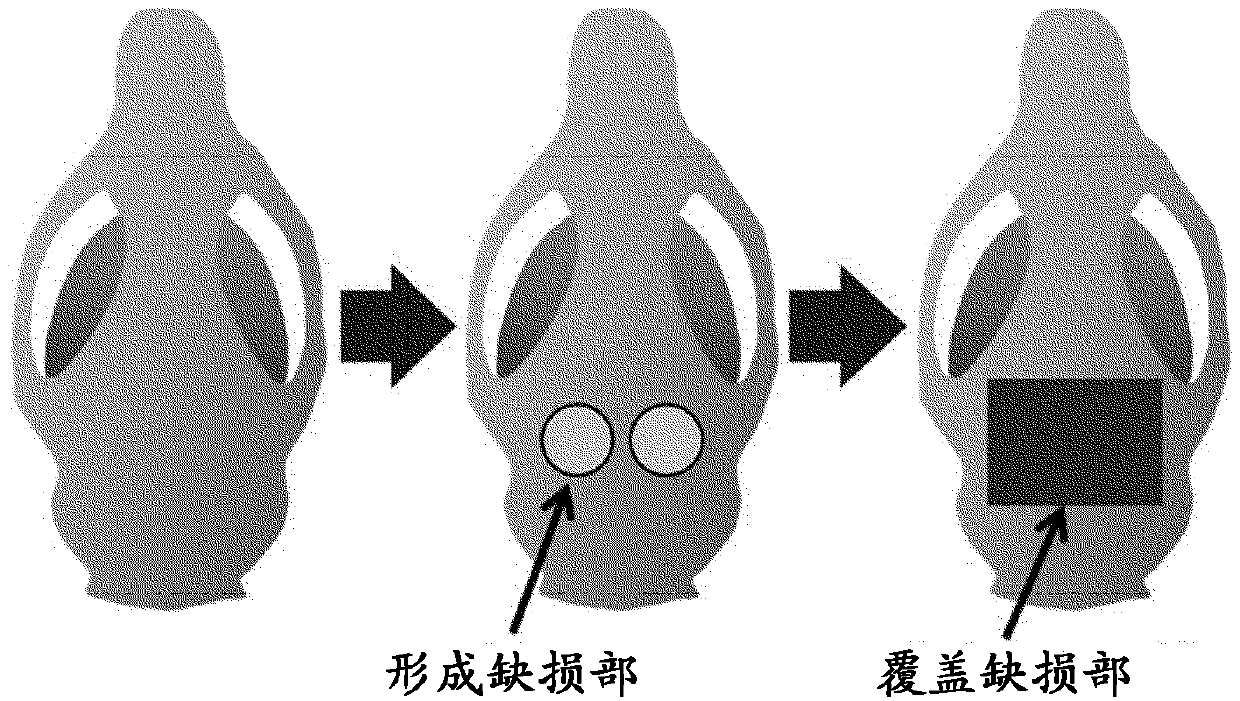
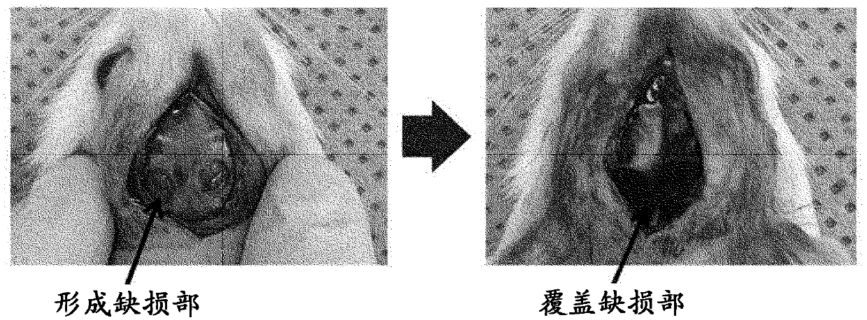
[0178] Nine-week-old LEW rats were generally anesthetized with 2 to 3% isoflurane, and then infiltrated anesthetized with lidocaine containing 1 / 10,000 epinephrine in the surgical area. After hair removal in an area wider than the incision, a straight incision is made on the top of the head to the subperiosteum so that the surgical area is clearly visible. After the skin-periosteal flap was peeled off to expose the cranium, two circular bone defects with a diameter of 4 mm were formed by injecting sterile saline using a Trefin rod. After the defect was covered with a sterilized porous polyimide membrane, the skin and periosteum flap was reset and sutured with nylon thread. At this time, a model in which the mesh surface (side A) of the porous polyimide membrane was placed in contact with the wound and a model in which the large-pore surface (side B) was placed in contact with the wound were produced. The healing status of the defect was measured after 2 weeks, 4 weeks, and 8 ...

Embodiment 2

[0180] Femur and neck bones were collected from 8-week-old GFP transgenic rats, both ends of the respective bones were cut, and bone marrow cell masses were collected by washing with DMEM medium supplemented with 10% FBS. The cell mass was pulverized by pipetting, and 1.0×10 6Bone marrow cells were statically cultured in DMEM medium supplemented with 10% FBS for 5 days. On day 6, the porous polyimide membrane to which the cells were attached was washed with a phosphate buffer, the medium was replaced with DMEM only, and cultured for another day. Nine-week-old nude mice were generally anesthetized with 2 to 3% isoflurane, and then infiltrated anesthetized with lidocaine containing 1 / 10,000 epinephrine in the surgical area. After hair removal in an area wider than the incision, a straight incision is made on the top of the head to the subperiosteum so that the surgical area is clearly visible. After the skin-periosteal flap was peeled off to expose the cranium, two circular bo...

Embodiment 3

[0182] Femur and neck bones were collected from 8-week-old GFP transgenic rats, both ends of the respective bones were cut, and bone marrow cell masses were collected by washing with DMEM medium supplemented with 10% FBS. The cell mass was pulverized by pipetting, and 1.0×10 6 Bone marrow cells were statically cultured in DMEM medium supplemented with 10% FBS for 5 days. After replacing the DMEM medium, culture was carried out for another 1 day. Then, inoculate 1.0×10 6 Bone marrow cells were statically cultured in DMEM medium supplemented with 10% FBS for 5 days. On day 6, the porous polyimide membrane to which the cells were attached was washed with a phosphate buffer, the medium was replaced with DMEM only, and cultured for another day. With respect to the obtained porous polyimide membrane, 9-week-old nude mice were generally anesthetized with 2 to 3% isoflurane, and then infiltrated anesthetized with lidocaine containing 1 / 10,000 epinephrine in the surgical area. Afte...


PUM

PropertyMeasurementUnit
pore sizeaaaaaaaaaa
pore sizeaaaaaaaaaa
thicknessaaaaaaaaaa

Abstract

The present invention pertains to an implant for treatment of a bone lesion site, the implant including a polymer porous film and a layer containing a biocompatible material, wherein the polymer porous film is a polymer porous membrane of a three-layer structure having a porous surface layer A, a porous surface layer B, and a macrovoid layer interposed between the surface layer A and the surface layer B; the average pore diameter of the pores in surface layer A is smaller than the average pore diameter of the pores in surface layer B; the macrovoid layer has a partition joined to the surface layers A and B and a plurality of macrovoids enclosed by the partition and the surface layers A and B; and the pores in the surface layers A and B communicate with the macrovoids.

Description

technical field [0001] The present invention relates to an implant and a kit for treating a bone injury site, and a treatment method for a bone injury site. Background technique [0002] Bone Injury and Healing [0003] From the viewpoint of patient QOL, healing of bone injuries including fractures and bone defects has been tried since ancient times by methods such as fixing the affected part with plates or screws, and surgical methods using a large number of instruments and devices. In recent years, bone disease treatment materials and devices having various structural features, functional features, and biocompatibility features have been created and used in medicine (Non-Patent Document 1, Patent Document 1). For example, titanium mesh can be used for the treatment of many bone injuries and oral diseases due to its high firmness, flexibility and adaptability to life. Titanium mesh can provide a stable wound healing space, but it does not have a bone healing effect by it...

Claims


Application Information

Patent Timeline
Patent Type & Authority Applications(China)
IPC IPC(8): A61L27/18A61K35/28A61K35/32A61K35/33A61L27/06A61L27/12A61L27/22A61L27/24A61L27/38A61L27/44A61L27/56
CPCA61L27/06A61L27/12A61L27/18A61L27/22A61L27/24A61L27/38A61L27/44A61L27/56A61K35/28A61K35/32A61K35/33A61L27/3821A61L27/3847A61L2430/02A61F2/28A61F2/2846A61F2002/30784A61P19/00B32B2307/402B32B27/285B32B15/08B32B2255/26B32B27/08B32B2307/732B32B15/02B32B27/281B32B2255/10B32B3/26B32B2535/00B32B15/18B32B27/286C08L79/08C08L89/06C08L81/06A61F2/0077A61L27/3804A61F2/30767A61F2/4603A61F2002/30769A61F2002/30909A61L27/26A61L27/34A61L27/3834
Inventor 萩原昌彦太田启介平岛伸悟
Owner UBE IND LTD
Why Eureka
Try Eureka