Ultrasonic imaging method and device, and storage medium

An ultrasonic imaging method and ultrasonic imaging technology, which are applied in the directions of ultrasonic/sonic/infrasonic equipment control, ultrasonic/sonic/infrasonic diagnosis, ultrasonic/sonic/infrasonic Permian technology, etc., which can solve the problems of low detection efficiency, etc.

Active Publication Date: 2019-04-05
SHENZHEN MINDRAY BIO MEDICAL ELECTRONICS CO LTD +1
View PDF20 Cites 6 Cited by
  • Summary
  • Abstract
  • Description
  • Claims
  • Application Information

AI Technical Summary

Problems solved by technology

[0004] However, the detection methods in traditional technologies have low detection efficiency

Method used


Image

  • Ultrasonic imaging method and device, and storage medium
  • Ultrasonic imaging method and device, and storage medium
  • Ultrasonic imaging method and device, and storage medium

Examples


Embodiment Construction

[0056] In order to make the purpose, technical solution and advantages of the present application clearer, the present application will be further described in detail below in conjunction with the accompanying drawings and embodiments. It should be understood that the specific embodiments described here are only used to explain the present application, and are not intended to limit the present application.

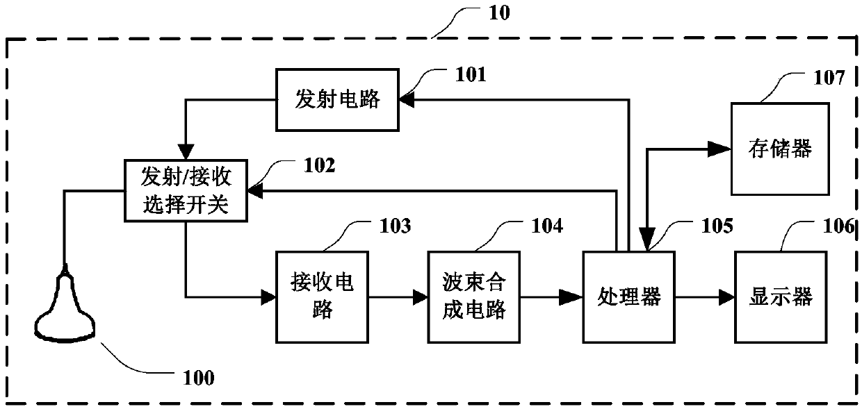
[0057] The ultrasonic imaging method provided in the embodiment of the present application can be applied to figure 1 In the ultrasound imaging device shown, figure 1 It is a schematic structural block diagram of the ultrasonic imaging device 10 in the embodiment of the present application. The ultrasonic imaging device 10 may include a probe 100 , a transmitting circuit 101 , a transmitting / receiving selection switch 102 , a receiving circuit 103 , a beam forming circuit 104 , a processor 105 and a display 106 . The transmitting circuit 101 can stimulate the probe 100 t...


PUM


Abstract

The invention relates to an ultrasonic imaging method and device, and a storage medium. The ultrasonic imaging method includes the steps that check parameters corresponding to a default check mode areadopted to perform the ultrasonic imaging process, a trigger instruction of a first section plane scanning check protocol is received, the first section plane scanning check protocol comprises one ormore pieces of section plane identification, the section plane scanning check process is started according to the first section plane scanning check protocol, and the section plane scanning check process comprises the steps that first target section plane identification is determined from the one or more pieces of section plane identification, according to the corresponding relationship between preset section plane identification and a check mode, a first check mode corresponding to the first target section plane identification is determined, and the ultrasonic imaging process is performed according to check parameters corresponding to the first check mode. By adopting the ultrasonic imaging method, the check efficiency can be improved, and the accuracy of checking images and check results is improved.

Description

technical field [0001] The present application relates to the technical field of medical equipment, in particular to an ultrasonic imaging method, equipment and storage medium. Background technique [0002] With the rapid development of the field of medical equipment, the auxiliary role of medical equipment is increasingly inseparable from clinical practice. As a common imaging device, ultrasonography equipment occupies an important position in clinical use. [0003] Usually, during an ultrasound examination, a doctor needs to perform an examination on multiple parts of an object to be detected, so as to determine the examination results of each part according to the section image of each part. However, since the characteristics of each part to be inspected are different, in order to obtain a section image of each part to be inspected, it is often necessary for the doctor to manually set the inspection parameters of the inspection equipment for different parts to be inspect...

Claims


Application Information

Patent Timeline
Patent Type & Authority Applications(China)
IPC IPC(8): A61B8/00
CPCA61B8/48A61B8/54A61B8/585
Inventor 姜玉新温博孟华王红燕徐钟慧
Owner SHENZHEN MINDRAY BIO MEDICAL ELECTRONICS CO LTD
Why Eureka
Try Eureka