Mammary gland lesion area detection method based on deep learning and transfer learning

A technology of transfer learning and deep learning, applied in the direction of neural learning methods, instruments, biological neural network models, etc., can solve problems such as unconsidered, high false positive rate, complexity of a large number of parameter adjustment processes, etc., and achieve the effect of improving the prediction effect

Pending Publication Date: 2019-04-16
深圳蓝影医学科技股份有限公司
View PDF11 Cites 43 Cited by
  • Summary
  • Abstract
  • Description
  • Claims
  • Application Information

AI Technical Summary

Problems solved by technology

The disadvantage of this method is that only the gray value of the image block is used for analysis, and the texture, edge, shape and other information of the image are not considered, which will affect the detection rate of the tumor to a certain extent, and will also cause high false positives. Rate
The shortcoming

Method used


Image

  • Mammary gland lesion area detection method based on deep learning and transfer learning
  • Mammary gland lesion area detection method based on deep learning and transfer learning
  • Mammary gland lesion area detection method based on deep learning and transfer learning

Examples


Embodiment 1

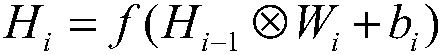
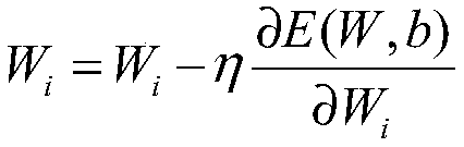
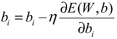
[0059] Example 1: Preparation and amplification of training set and test set; According to the tumor location information marked by doctors in the breast data set, the available tumor image is extracted and its size is normalized to 100*100 pixel size as a positive sample. Randomly determine the same amount of normal tissue with a size of 100*100 pixels on the mammary gland image as a negative sample. The positive and negative samples are rotated 90, 180, 270 degrees and flipped up and down, left and right, so that the final training data contains 840 equal positive and negative samples. The class standard of positive samples is set to 1, and the class standard of negative samples is set to 0;

[0060] The target image block is prepared; the original breast image is down-sampled, and the breast contour is obtained by using the maximum inter-class variance method to determine the maximum range of the effective breast area; Slide from left to right and from top to bottom to obt...


PUM


Abstract

The invention provides a mammary gland lesion area detection method based on deep learning and transfer learning. The method comprises: preparation and amplification of a training set and a test set;according to lump position information marked by a doctor in the breast data set, extracting an available lump image and normalizing the size of the available lump image into a size of 100 * 100 pixels as a positive sample; according to the invention, the AlexNet network is used to train the parameter model of the classification model of the natural image on the ImageNet data set; training and transfer learning are carried out on a specific breast image data set, so that the binary classification problem of the convolutional neural network on a small-scale breast data set can be successfully solved, a lesion area in the breast image can be identified, and the prediction effect on the breast lesion is improved.

Description

technical field [0001] The invention is a method for detecting breast lesion areas based on deep learning and transfer learning, which belongs to the medical field. Background technique [0002] In the prior art, breast cancer is a common malignant tumor, and early diagnosis and treatment are the key to reducing breast cancer mortality. Lesions in breast images include masses, calcifications, bilateral asymmetry, and structural distortions. Masses and calcification clusters are the most common imaging signs of breast cancer. Therefore, automatic detection of masses and calcifications has also become a computer-aided method. There are two main aspects of the diagnostic system. Among them, due to factors such as blurred edges, various shapes, and low contrast with surrounding tissues, tumors have always been a major difficulty in computer-aided detection. Therefore, if not emphasized, the lesion area in this patent all refers to lump. [0003] The idea of ​​deep learning or...

Claims


Application Information

Patent Timeline
IPC IPC(8): G06K9/62G06N3/04G06N3/08
CPCG06N3/08G06N3/084G06N3/045G06F18/2411G06F18/214
Inventor 胡阳郭朋郑杰陈晶鄢照龙
Owner 深圳蓝影医学科技股份有限公司
Why Eureka
Try Eureka